. 2002 Dec;88(6):604-10.
doi: 10.1136/heart.88.6.604.

Antiarrhythmic and anti-ischaemic effects of angina in patients with and without coronary collaterals

Affiliations

Antiarrhythmic and anti-ischaemic effects of angina in patients with and without coronary collaterals

R J Edwards et al. Heart. 2002 Dec.

Abstract

Objective: To determine whether the changes in the manifestations of myocardial ischaemia during sequential angina episodes caused by exercise or coronary artery occlusion are collateral dependent.

Methods: 40 patients awaiting percutaneous transluminal coronary angioplasty for an isolated left anterior descending artery stenosis underwent three sequential treadmill exercise tests, with the second exertion separated from the first by 15 minutes, and from the third by 90 minutes; 28 patients subsequently completed two (> 180 s) sequential intracoronary balloon inflations with measurement of collateral flow index from mean coronary artery wedge, aortic, and coronary sinus pressures.

Results: On second compared with first exercise, time to 0.1 mV ST depression (mean (SD): 340 (27) v 266 (25) s) and rate-pressure product at 0.1 mV ST depression (22 068 (725) v 19 586 (584) beats/min/mm Hg) were increased (all p < 0.005), while angina and ventricular ectopic beat frequency were diminished (p < 0.05). This advantage, which had waned by the third effort, was independent of collateral flow index. Similarly, at the end of the second compared with the first coronary occlusion, ventricular tachycardia (21% v 0%, p < 0.05), ST elevation (0.47 (0.07) v 0.33 (0.05) mV, p < 0.005), and angina severity (6.1 (0.7) v 4.6 (0.7) units, p < 0.005) were reduced despite similar collateral flow indices.

Conclusions: In patients with coronary artery disease, ventricular arrhythmias, ST deviation, and angina are reduced during a second exertion or during a second coronary occlusion. This protective effect can occur independently of collateral recruitment. These characteristics, together with the breadth and temporal pattern of protection, are consistent with ischaemic preconditioning.


Figures
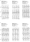
Figure 1
Figure 1
The appearance of the surface ECG during repetitive exercise. Patients were subjected to three consecutive treadmill exercise tests. Test 2 started 15 minutes after test 1 had been completed, and test 3 was begun 90 minutes after the end of test 2. Panels A and B consist of serial ECG recordings taken at identical time points into each of the three exercise tests in two different patients. (A) ST segment depression occurs in tests 1 and 3, but is absent at the identical time point in test 2 despite a similar rate–pressure product. (B) The record of a patient who developed ventricular bigeminy during tests 1 and 3. During these tests, the complexes with a narrow QRS morphology represent normally conducted beats with 0.4 mV of ST segment depression. At the corresponding time point during test 2, ST segment depression is less pronounced (0.2 mV) and is no longer accompanied by ventricular bigeminy.
Figure 2
Figure 2
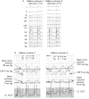
The effect of serial coronary artery occlusions on the surface ECG, intracoronary ECG, and pressures determining myocardial collateral blood flow. (A) The appearance of the surface ECG after 180 seconds of occlusion of the left anterior descending coronary artery (LAD) during percutaneous transluminal coronary angioplasty. The second occlusion began five minutes after the end of the first occlusion. The first episode of ischaemia causes ST segment elevation and QRS broadening without a shift in R wave axis. These changes are much less pronounced during the second occlusion. ST segment elevation confirms the presence of transmural myocardial ischaemia during both occlusions. (B) Records of pressures and intracoronary ECG during serial coronary artery occlusion. These traces were synchronous with the surface ECG appearing in panel A. The lowermost trace represents the intracoronary ECG recorded from the angioplasty guide wire. The intracoronary ECG reflects the changes seen in panel A with pronounced attenuation of ST segment elevation on second occlusion. The next uppermost trace is the pressure in the LAD distal to the site of balloon occlusion. The next uppermost trace is of the coronary sinus pressure (CSP) which is on a 25 mm Hg scale, as opposed to a 100 mm scale. Flow across the myocardial bed is largely determined by the difference between the venous and arterial pressure. During the first and second balloon occlusions the mean distal arterial pressure is virtually the same as the mean coronary sinus pressure and therefore there is no appreciable gradient to drive myocardial blood flow. Despite the absence of a change in myocardial blood flow, myocardial ischaemia is notably attenuated during the second inflation. This implies that myocardial resistance to ischaemia is enhanced during the second coronary artery occlusion. (For an explanation of the relation between pressure and collateral flow, see Methods.)
Figure 3
Figure 3
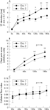
Changes in (A) ST segment deviation, (B) chest pain intensity, and (C) collateral flow index during the first and second coronary artery occlusions. Chest discomfort and ST segment deviation are significantly reduced on the second compared with the first balloon occlusion. In keeping with the changes seen in fig 2, the reduction in these measures of myocardial ischaemia is not accompanied by an increase in myocardial collateral blood flow. In patients reaching three minutes of balloon occlusion the collateral flow increased in both groups with increasing duration of ischaemia. *p < 0.05, occlusion 1 v occlusion 2; §p < 0.005, occlusion 1 v occlusion 2; †p < 0.001, occlusion 1 v occlusion 2; ‡p < 0.0005 occlusion 1 v occlusion 2.
Figure 4
Figure 4
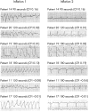
Cardiac rhythm at peak inflation in patients with ventricular tachycardia associated with coronary artery occlusion. The left panel shows traces from patients developing ventricular arrhythmias during the first intracoronary balloon occlusion. The right panel shows traces at similar time points during the second occlusion. Note that ventricular tachycardia was confined to first balloon occlusion and absent during the second at the same and later time points. The collateral flow index at the time of recording appears above each trace. Collateral flow indices below 0.25 are thought insufficient to prevent ischaemia.

References

    1. Heberden J. Letter to the Editor. Medical Transactions, College of Physicians London 1785;3:1–11.
    1. Marber MS, Joy MD, Yellon DM. Warm-up in angina: is it ischaemic preconditioning? Br Heart J 1994;72:213–15. - PMC - PubMed
    1. Yellon DM, Baxter GF, Marber MS. Angina reassessed: pain or protector? Lancet 1996;347:1159–62. - PubMed
    1. Murry CE, Jennings RB, Reimer KA. Preconditioning with ischaemia: a delay of lethal cell injury in ischemic myocardium. Circulation 1986;74:1124–36. - PubMed
    1. Kloner RA, Bolli R, Marban E, et al. Medical and cellular implications of stunning, hibernation, and preconditioning: an NHLBI workshop. Circulation 1998;97:1848–67. - PubMed

Publication types

MeSH terms