. 2002 Nov 15;545(1):207-16.
doi: 10.1113/jphysiol.2002.021980.

Secretion and cell volume regulation by salivary acinar cells from mice lacking expression of the Clcn3 Cl- channel gene

Affiliations

Secretion and cell volume regulation by salivary acinar cells from mice lacking expression of the Clcn3 Cl- channel gene

Jorge Arreola et al. J Physiol. .

Abstract

Salivary gland acinar cells shrink when Cl(-) currents are activated following cell swelling induced by exposure to a hypotonic solution or in response to calcium-mobilizing agonists. The molecular identity of the Cl(-) channel(s) in salivary cells involved in these processes is unknown, although ClC-3 has been implicated in several tissues as a cell-volume-sensitive Cl(-) channel. We found that cells isolated from mice with targeted disruption of the Clcn3 gene undergo regulatory volume decrease in a fashion similar to cells from wild-type littermates. Consistent with a normal regulatory volume decrease response, the magnitude and the kinetics of the swell-activated Cl(-) currents in cells from ClC-3-deficient mice were equivalent to those from wild-type mice. It has also been suggested that ClC-3 is activated by Ca(2+)-calmodulin-dependent protein kinase II; however, the magnitude of the Ca(2+)-dependent Cl(-) current was unchanged in the Clcn3(-/-) animals. In addition, we observed that ClC-3 appeared to be highly expressed in the smooth muscle cells of glandular blood vessels, suggesting a potential role for this channel in saliva production by regulating blood flow, yet the volume and ionic compositions of in vivo stimulated saliva from wild-type and null mutant animals were comparable. Finally, in some cells ClC-3 is an intracellular channel that is thought to be involved in vesicular acidification and secretion. Nevertheless, the protein content of saliva was unchanged in Clcn3(-/-) mice. Our results demonstrate that the ClC-3 Cl(-) channel is not a major regulator of acinar cell volume, nor is it essential for determining the secretion rate and composition of saliva.


Figures

Figure 1
Figure 1. ClC-3 protein and mRNA expression in mouse parotid acinar cells
Protein was isolated from the parotid glands of ClC-3 wild-type and null mutant mice. Upper panel, an antibody to ClC-3 recognized several bands of the appropriate molecular mass for ClC-3 in protein samples from a wild-type mouse parotid gland (lane 1). In a protein sample from parotid glands of Clcn3−/-mice, a band of the correct molecular mass for ClC-3 protein was not observed (lane 2), although several other bands remained immunoreactive. These most likely represent cross-reactivity of the antibody with non-specific proteins. When probed with an anti-ClC-3 antibody that had been pre-absorbed with a threefold excess of the peptide antigen to which the antibody had been raised (lane 3) or with secondary antibody alone (lane 4), none of the immunoreactive bands in the wild-type protein sample were recognized. Molecular weight markers are shown to the right of the blots. Lower panel, four overlapping RT-PCR primers were designed to amplify the ClC-3 message from isolated parotid acinar cells including two sense primers (U1 and U2) complimentary to codons 34–40 and 384-391, respectively, and two nonsense primers corresponding to codons 209–214 and 590–597 (L1 and L2, respectively). The PCR product sizes using primer pairs U1/L1 and U2/L2 are 542 base pairs (bp) and 638 bp long, respectively. DNA sequencing confirmed 100 % identity of the PCR products as ClC-3. Lane M is a 100 bp DNA ladder size marker.
Figure 2
Figure 2. Immunohistochemical labelling using a ClC-3 antibody in the parotid glands of Clcn3+/+and Clcn3−/-mice
The parotid glands were removed from Clcn3+/+ and Clcn3−/- mice and fixed in paraformaldehyde overnight prior to imbedding in paraffin and sectioning at 5 μm. The glands were exposed to anti-ClC-3 antibody (1:250) overnight. Reactivity was then detected by the use of an Alexa-594 fluorescent secondary antibody. Images were taken at several magnifications from × 200 to × 600, as indicated below. Upper left panel, section from wild-type parotid gland. Arrows indicate intense staining of blood vessels (magnification, ×200). Upper right panel, section of parotid gland from null mutant mouse. Intense staining of blood vessels is absent, but non-specific staining of duct and acinar cells remains (magnification, ×200). Lower left panel, section from wild-type parotid gland. Arrows indicate intense staining of blood vessels (magnification, ×600). Lower right panel, Nomarski image of the section in the lower left panel (magnification, ×600).
Figure 3
Figure 3. Targeted disruption of the Clcn3 gene fails to inhibit the RVD response in parotid acinar cells
The role of ClC-3 in the regulatory volume decrease (RVD) response was examined in parotid acinar cells loaded with the fluoroprobe calcein, as described in Methods. Upper panel, parotid acini isolated from Clcn3+/+ (▪) and Clcn3−/- (▵) mice were superfused in an isosmotic solution and then hyposomotic cell swelling was induced by switching the perfusate to a hypotonic medium (30 % dilution with water). The cell volumes shown are normalized to the maximum volume achieved following exposure to a hypotonic solution. Changes in cell volume are represented as 1/normalized calcein fluorescence (1/calcein Fn). Lower panel, the initial rate of change expressed as 1/calcein Fn × min−1 × 10⁻² (Clcn3+/+, n = 8; Clcn3/-, n = 11; no statistical differences between groups).
Figure 4
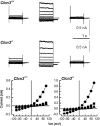
Figure 4. Swell-activated Cl currents in parotid acinar cells from Clcn3+/+ and Clcn3/- mice
Volume-sensitive Cl currents recorded from acinar cells isolated from Clcn3+/+ (upper row) and Clcn3−/- (middle row) mice. Control traces (left column) were recorded after 5 min of dialysis under isotonic conditions; hypotonic-activated currents (centre column) were recorded after 2–3 min in the hypotonic solution; inhibition of Cl currents (right column) by cell shrinkage was assessed after 5 min exposure to a hypertonic solution. The lower row shows the current-voltage relationship for the data depicted in the upper and middle rows under isotonic (▪), hypotonic (•) and hypertonic (▴) conditions obtained from acinar cells isolated from Clcn3+/+ and Clcn3/- mice. There was no statistical difference between the swell-activated current (current present in the hypotonic solution minus the current after switching to the hypertonic solution) from wild-type and null mutant mice (Clcn3+/+ = 29.2 ± 8.2 pA pF−1, n = 6; Clcn3/- = 32.2 ± 3.9 pA pF−1, n = 7).
Figure 5
Figure 5. Ca2+-dependent Cl currents in parotid acinar cells from Clcn3+/+and Clcn3−/-mice
Upper panel, currents from a parotid acinar cell from a Clcn3+/+ mouse in response to 3 s voltages pulses from −30 to +90 mV in 20 mV increments from a holding potential of −50 mV. Lower panel, currents at the same potentials from a parotid acinar cell from a Clcn3−/- mouse. There was no statistical difference between the Ca2+-activated current from wild-type and null mutant mice (Clcn3+/+, 140 ± 20 pA pF−1; n = 10; Clcn3−/-, 135 ± 13 pA pF−1; n = 5).
Figure 6
Figure 6. Targeted disruption of the Clcn3 gene does not inhibit pilocarpine-induced in vivo salivation
The role of ClC-3 in the production of saliva was examined in vivo by stimulating with 10 mg pilocarpine-HCl (kg body weight (BW))−1. Whole saliva was collected from the lower cheek pouch by suctioning at intervals of 5, 10 and 15 min and amounts are expressed as μl 5 min g BW−1. Time zero was designated as the point when saliva was first noted following pilocarpine injection. Clcn3+/+ (▪), n = 17; Clcn3−/- (□), n = 12.

References

    1. Arreola J, Melvin JE, Begenisich T. Activation of calcium-dependent chloride channels in rat parotid acinar cells. Journal of General Physiology. 1995;108:35–47. - PMC - PubMed
    1. Arreola J, Melvin JE, Begenisich T. Volume-activated chloride channels in rat parotid acinar cells. Journal of Physiology. 1996;484:677–687. - PMC - PubMed
    1. Begenisich T, Melvin JE. Regulation of chloride channels in secretory epithelia. Journal of Membrane Biology. 1998;163:77–85. - PubMed
    1. Bers DM, Patton CW, Nuccitelli R. A practical guide to the preparation of Ca2+ buffers. Methods in Cell Biology. 1994;40:3–29. - PubMed
    1. Castle JD, Cameron RS, Arvan P, Von Zastrow M, Rudnick G. Similarities and differences among neuroendocrine, exocrine, and endocytic vesicles. Annals of the New York Academy of Sciences. 1987;493:448–460. - PubMed

Publication types

LinkOut - more resources