. 2002 Nov 15;545(1):321-30.
doi: 10.1113/jphysiol.2002.018010.

Force enhancement following muscle stretch of electrically stimulated and voluntarily activated human adductor pollicis

Affiliations

Force enhancement following muscle stretch of electrically stimulated and voluntarily activated human adductor pollicis

Hae-Dong Lee et al. J Physiol. .

Abstract

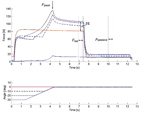
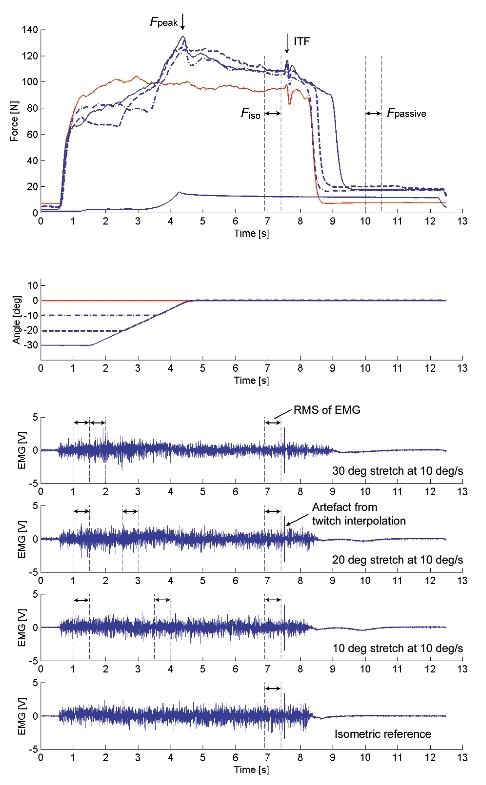
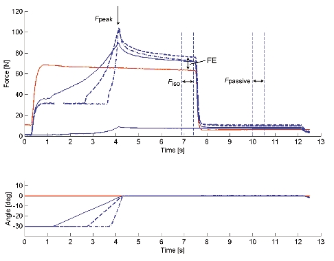
For electrically stimulated muscles, it has been observed that maximal muscle force during and after stretch is substantially greater than the corresponding isometric force. However, this observation has not been made for human voluntary contractions. We investigated the effects of active muscle stretch on muscle force production for in vivo human adductor pollicis (n = 12) during maximal voluntary contractions and electrically induced contractions. Peak forces during stretch, steady-state isometric forces following stretch, and passive forces following muscle deactivation were compared to the corresponding isometric forces obtained at optimal muscle length. Contractions with different stretch magnitudes (10, 20, and 30 deg at a constant speed of 10 deg s(-1)) and different speeds (10, 20, and 60 deg s(-1) over a range of 30 deg) were performed in triplicate in a random order, balanced design. We found three novel results: (i) there was steady-state force enhancement following stretch in voluntarily contracted muscles; (ii) some force enhancement persisted following relaxation of the muscle and (iii) force enhancement, for some stretch conditions, exceeded the maximum isometric force at optimal muscle length. We conclude from these results that voluntary muscle contraction produces similar force enhancement to that observed in the past with electrically stimulated preparations. Therefore, steady-state force enhancement may play a role in everyday movements. Furthermore, these results suggest that non-uniformities in sarcomere length do not, at least not exclusively, account for the force enhancement following active muscle stretch, and that the stretch magnitude-dependent passive force enhancement observed here may be responsible for the enhancement of force above the isometric reference force at optimal muscle length.


Figures

Figure 1
Figure 1. Testing apparatus and experimental set up
The testing apparatus used consists of a rotary stepper motor (a) with a digital stepper drive/controller (b), an aluminium rod (c) instrumented with two pairs of strain gauges (d) and an auxiliary piece for thumb placement (e), with an analog encoder (f). When the adductor pollicis contracts, the thumb presses against the rod and thumb adduction force is measured by the calibrated strain gauges. The forearm and the four digits of the left hand are restrained with a clinical cast (g) and secured on the apparatus using velcro straps. The thumb is placed and aligned on the rod as shown in the pictures. This experimental set up was designed to measure thumb adduction force during voluntary and electrically elicited, static and dynamic contractions.
Figure 2
Figure 2. Thumb adduction force and angle - time histories of test and reference contractions obtained for Protocol 1 during electrical stimulation
For the test contractions, peak force (Fpeak) was measured during the stretch phase. Isometric force (Fiso) was measured during the final, steady-state isometric phase for comparisons with the corresponding values obtained from the isometric reference contractions and used to assess force enhancement (FE). After deactivation of the muscle, passive force (Fpassive) was measured. Also, purely passive forces were measured for stretch contractions of the deactivated muscle (a stretch contraction over 30 deg stretch at 10 deg s−1 is presented).
Figure 3
Figure 3. Thumb adduction force and angle - time histories and EMG signals of test and reference contractions obtained for Protocol 1 during voluntary contraction
For the test contractions, peak force (Fpeak) was measured during the stretch phase, and isometric force (Fiso), EMG, and interpolated twitch force (ITF) were measured during the final isometric phase for comparisons with the corresponding values obtained from the isometric reference contractions. After deactivation of the muscle, passive force (Fpassive) was measured. Also, purely passive forces were measured for stretch contractions of the completely relaxed muscle (a stretch contraction over 30 deg stretch at 10 deg s−1 is presented).
Figure 4
Figure 4. Thumb adduction force and angle - time histories of test and reference contractions obtained for Protocol 2 during electrical stimulation
For the test contractions, peak force (Fpeak) was measured during the stretch phase. Isometric force (Fiso) was assessed during the final, steady-state isometric phase for comparisons with the corresponding values obtained from the isometric reference contractions and used to assess force enhancement (FE). After deactivation of the muscle, passive force (Fpassive) was measured. Also, purely passive forces were measured for stretch contractions of the deactivated muscle (a stretch contraction over 30 deg stretch at 10 deg s−1 is presented).
Figure 5
Figure 5. Thumb adduction force and angle - time histories and EMG signals of test and reference contractions obtained for Protocol 2 during voluntary contraction
For the test contractions, peak force (Fpeak) was measured during the stretch phase, and isometric force (Fiso), EMG, and interpolated twitch force (ITF) were measured during the final isometric phase for the comparison with the corresponding values obtained from the isometric reference contractions. After deactivation of the muscle, passive force (Fpassive) was measured. Also, purely passive forces were measured for stretch contractions of the completely relaxed muscle (a stretch contraction over 30 deg stretch at 10 deg s−1 is presented).
Figure 6
Figure 6. Peak force reached during muscle stretch (Fpeak) for different conditions
All peak forces were normalized relative to the corresponding isometric reference forces and pooled for each stretch condition and across all subjects. A, Protocol 1: the peak forces during stretch were greater for stretch amplitudes of 20 and 30 deg than those for 10 deg during electrical stimulation. Fpeak was found to be greatest for stretch amplitudes of 20 deg for the voluntary contractions. Significance (* P < 0.05) is presented separately for electrically induced (continuous line) and for voluntary (dashed line) contractions because a significant interaction (stimulation method × stretch condition) was found. B, Protocol 2: Fpeak increased with increasing speeds of stretch for the electrical and voluntary contractions. Means were collapsed across the stimulation methods and significance (* P < 0.05) is presented only for different stretch conditions, as no interaction (stimulation method × stretch condition) was found. Fpeak, peak force produced during active stretching.
Figure 7
Figure 7. Steady-state force enhancement (FE) following muscle stretch
Steady-state force enhancement values were pooled for each stretch condition and across all subjects. A, Protocol 1: the steady-state force enhancement following 20 and 30 deg stretches was significantly greater than that following 10 deg stretches for the tests using electrical stimulation. Force enhancement was greatest following 20 deg of stretch for the voluntary contractions. Significance (* P < 0.05) is presented separately for electrically induced (continuous line) and for voluntary (dashed line) contractions because a significant interaction (stimulation method × stretch condition) was found. B, Protocol 2: force enhancement was not influenced by the speed of stretch regardless of the method of stimulation. FE: force enhancement following muscle stretching.
Figure 8
Figure 8. Passive force (Fpassive) assessed after the termination of stimulation
Passive force values were pooled for each stretch condition and across all subjects. A, Protocol 1: passive forces following the isometric reference contractions were significantly smaller than the passive forces following active muscle stretching († P < 0.05). There was no significant difference due to stimulation methods. Means were collapsed across the stimulation methods and significance (* P < 0.05) is presented only for different stretch conditions, as no interaction (stimulation method × stretch condition) was found. B, Protocol 2: passive forces following active muscle stretching were significantly greater than the passive forces following the isometric reference contractions († P < 0.05). There was no significant effect of the stimulation methods or the speed of stretching. Fpassive, passive force.
Figure 9
Figure 9. Muscle activation determined with EMG and the interpolated twitch technique during voluntary contractions
Muscle activation following active stretching was smaller than that during the isometric reference contractions as indicated by the smaller root mean square (RMS) values of the EMG and the greater interpolated twitch force (indicated with ITF/RTF) in the stretch tests compared to the reference contractions. Statistical significance (* P < 0.05) compared to the values of the isometric reference contractions. Muscle activation during active stretch was greater than just prior to stretch (†, P < 0.05). ITF, interpolated twitch force; RTF, resting twitch force; REF, isometric reference contraction, Before, RMS of EMG just before active stretch for 500 ms; During, RMS of EMG just during active stretch for 500 ms; After, RMS of EMG after active stretch for 500 ms during the steady-state isometric phase of the experimental stretch contraction.
Figure 10
Figure 10. The contributions of active and passive force enhancement to the total force enhancement (FE = FEactive + FEpassive)
The contribution of force enhancement observed during contractions was estimated based on the assumption that the passive component of force enhancement, as determined here, subtracted from the total force enhancement would give the contribution of an ‘active’ component (i.e. a component that disappears following deactivation of the muscle). FE, force enhancement following muscle stretching; FEactive, force enhancement following muscle stretching due to active components; FEpassive, force enhancement following muscle stretching due to passive components.

References

    1. Abbott BC, Aubert XM. The force exerted by active striated muscle during and after change of length. Journal of Physiology. 1952;117:77–86. - PMC - PubMed
    1. Belanger AY, McComas AJ. Extent of motor unit activation during effort. Journal of Applied Physiology. 1981;51:1131–1135. - PubMed
    1. Cook CS, McDonagh MJ. Force responses to controlled stretches of electrically stimulated human muscle-tendon complex. Experimental Physiology. 1995;80:477–490. - PubMed
    1. Crago PE, Houk JC, Hasan Z. Regulatory actions of human stretch reflex. Journal of Neurophysiology. 1976;39:925–935. - PubMed
    1. De Ruiter CJ, Didden WJM, Jones DA, De Haan A. The force-velocity relationship of human adductor pollicis muscle during stretch and the effects of fatigue. Journal of Physiology. 2000;526:671–681. - PMC - PubMed

Publication types

LinkOut - more resources