. 2002 Nov 15;30(22):4910-7.
doi: 10.1093/nar/gkf619.

Crystal structure of actinomycin D bound to the CTG triplet repeat sequences linked to neurological diseases

Affiliations

Crystal structure of actinomycin D bound to the CTG triplet repeat sequences linked to neurological diseases

Ming-Hon Hou et al. Nucleic Acids Res. .

Abstract

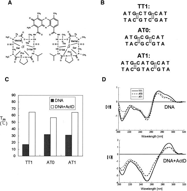
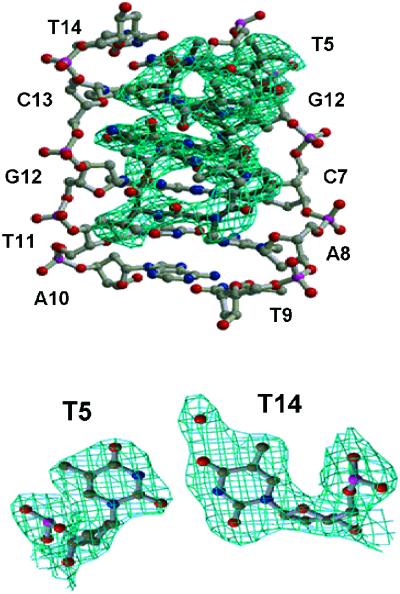
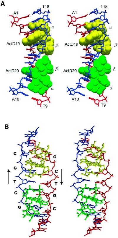
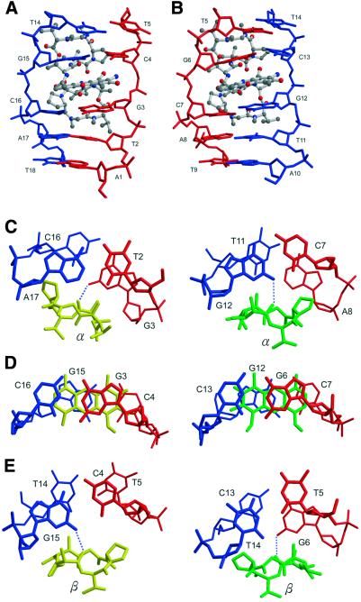
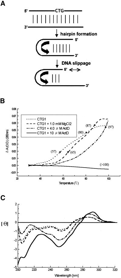
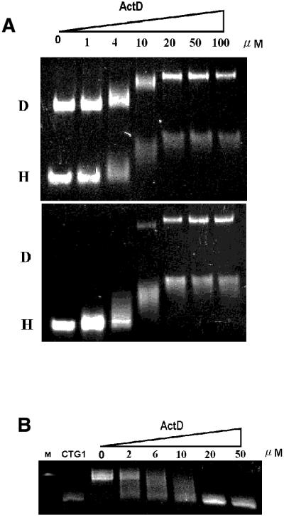
The potent anticancer drug actinomycin D (ActD) acts by binding to DNA GpC sequences, thereby interfering with essential biological processes including replication, transcription and topoisomerase. Certain neurological diseases are correlated with expansion of (CTG)n trinucleotide sequences, which contain many contiguous GpC sites separated by a single base pair. In order to characterize the binding of ActD to CTG triplet repeat sequences, we carried out heat denaturation and CD analyses, which showed that adjacent GpC sequences flanking a T:T mismatch are preferred ActD-binding sites, and that ActD binding results in a conformational transition to A-type structure. The structural basis of the strong binding of ActD to neighboring GpC sites flanking a T:T mismatch was provided by the crystal structure of ActD bound to ATGCTGCAT, which contains a CTG triplet sequence. Binding of two ActD molecules to GCTGC causes a kink in the DNA helix. In addition, using a synthetic self-priming DNA model, 5'-(CAG)4(CTG)(16)-3', we observed that ActD can trap the cruciform or duplexes of (CTG)n and interfere with the expansion process of CTG triplet repeats as shown by gel electrophoretic expansion assay. Our results may provide the possible biological consequence of ActD bound to CTG triplet repeat sequences.


Figures

Figure 1
Figure 1
(A) Chemical structure of ActD. The cyclic pentapeptide attached to the quinonoid ring and the benzenoid ring of the phenoxazone group are labeled α and β, respectively. (B) DNA duplexes that were used in this study include AT0, AT1 and TT1 (rectangles represent the binding sites for the phenoxazone ring of ActD). (C) The effect of ActD on the Tm values of AT0, AT1 and TT1 (4 µM) in standard buffer. (D) CD spectra of AT0, AT1 and TT1 (4 µM) in standard buffer alone (top) and with 10 µM ActD are graphed (bottom). The CD spectra of ActD–DNA complexes were obtained by subtracting the spectrum of ActD.
Figure 2
Figure 2
The refined (2FoFc) Fourier electron density map showing the five amino acids (threonine, N-methylvaline, sarcosine, proline and D-valine) and the T5·T14 mismatched base pair of the ActD–DNA complex at 2.6 Å resolution.
Figure 3
Figure 3
(A) Stereoscopic drawings of the crystal structure of the 2:1 ActD–(ATGCTGCAT)2 complex (ActD in van der Waals and DNA in skeletal representation). Two ActD molecules, ActD19 (yellow) and ActD20 (green), related by a two-fold symmetry operation, bind to DNA by intercalating the phenoxazone rings at the two GpC steps with cyclic pentapeptide moieties located in the minor groove. (B) Stereoscopic drawings of the ActD–DNA complex (ActD in ball-and-stick and DNA in skeletal line representation) viewed from the minor groove direction.
Figure 4
Figure 4
Close-up side view of the ActD–TGCAT part (ActD in ball-and-stick and DNA in skeletal representation). The two phenoxazone rings are intercalated individually into the (G3pC4)·(G15pG16) step in (A) and the (G6pC7)·(G12pC13) step in (B), respectively. (CE) The detailed conformation showing the stacking interactions in the ActD–DNA complex at various base pair steps of the refined structure. The hydrogen bonding between the oxygen atom of the threonine carbonyl group and the hydrogen atom of the guanine N2 amino group is marked by blue dotted lines.
Figure 5
Figure 5
(A) Scheme of hairpin formation and DNA slippage of CTG triplet repeats. (B) The melting profiles and Tm values of CTG1 (1 µM) in standard buffer and with ActD. (C) CD spectra of CTG1 (1 µM) in the same buffer solution. The CD spectra of ActD–DNA complexes were obtained by subtracting the spectrum of ActD alone.
Figure 6
Figure 6
(A) Gel mobility shift assay of CTG1 (1 µM) under different concentrations of ActD. D and H denote duplex and intrastrand hairpin forms of CTG1, respectively. (B) The interference assay of ActD on CTG1 (1 µM) expansion using DNA polymerase KF. The first (M) and second (CTG1) lanes represent the 90mer DNA marker and the 60mer (CAG)4(CTG)16 without polymerase, respectively. The other lanes show the 60mer extended with DNA polymerase in the presence of ActD.

References

    1. Sobell H.M. and Jain,S.C. (1972) Stereochemistry of actinomycin binding to DNA. II. Detailed molecular model of actinomycin-DNA complex and its implications. J. Mol. Biol., 68, 21–34. - PubMed
    1. Kamitori S. and Takusagawa,F. (1992) Crystal structure of the 2:1 complex between d(GAAGCTTC) and the anticancer drug actinomycin D. J. Mol. Biol., 225, 445–456. - PubMed
    1. Hurwitz J., Furth,J.J., Malamy,M. and Alexander,M. (1962) The role of deoxyribonucleic acid in ribonucleic acid synthesis. III: The inhibition of the enzymatic synthesis of ribonucleic acid and deoxyribonucleic acid by actinomycin D and proflavin. Proc. Natl Acad. Sci. USA, 48, 1222–1230. - PMC - PubMed
    1. Aivasashvilli V.A. and Beabealashvilli,R.S. (1983) Sequence-specific inhibition of RNA elongation by actinomycin D. FEBS Lett., 160, 124–128. - PubMed
    1. Chen F.M. (1988) Kinetic and equilibrium binding studies of actinomycin D with some d(TGCA)-containing dodecamers. Biochemistry, 27, 1843–1848. - PubMed

Publication types

MeSH terms

Associated data