. 2002 Nov 15;30(22):4975-84.
doi: 10.1093/nar/gkf630.

Novel repair activities of AlkA (3-methyladenine DNA glycosylase II) and endonuclease VIII for xanthine and oxanine, guanine lesions induced by nitric oxide and nitrous acid

Affiliations

Novel repair activities of AlkA (3-methyladenine DNA glycosylase II) and endonuclease VIII for xanthine and oxanine, guanine lesions induced by nitric oxide and nitrous acid

Hiroaki Terato et al. Nucleic Acids Res. .

Abstract

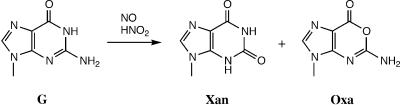
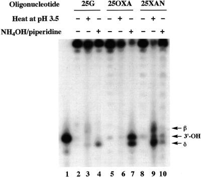
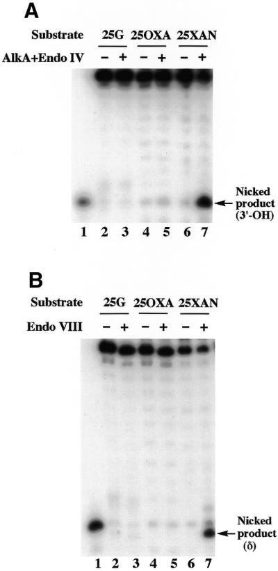
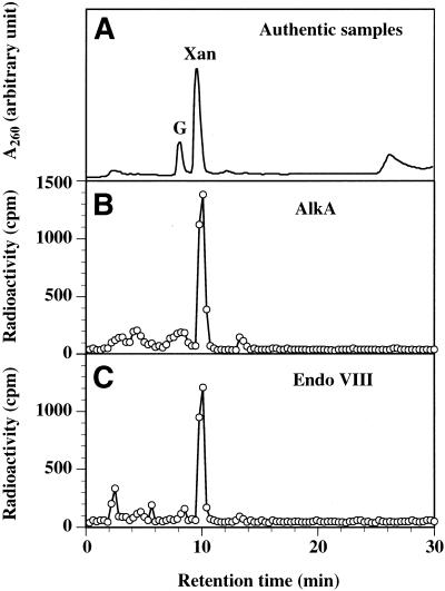
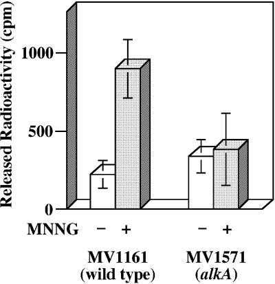
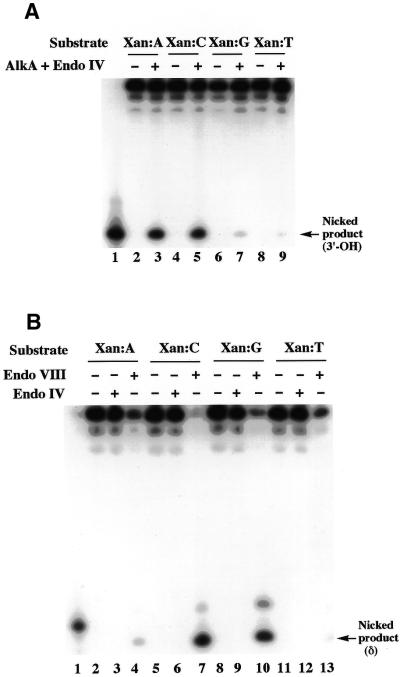
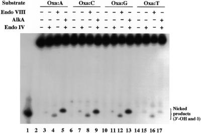
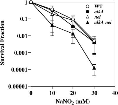
Nitrosation of guanine in DNA by nitrogen oxides such as nitric oxide (NO) and nitrous acid leads to formation of xanthine (Xan) and oxanine (Oxa), potentially cytotoxic and mutagenic lesions. In the present study, we have examined the repair capacity of DNA N-glycosylases from Escherichia coli for Xan and Oxa. The nicking assay with the defined substrates containing Xan and Oxa revealed that AlkA [in combination with endonuclease (Endo) IV] and Endo VIII recognized Xan in the tested enzymes. The activity (V(max)/K(m)) of AlkA for Xan was 5-fold lower than that for 7-methylguanine, and that of Endo VIII was 50-fold lower than that for thymine glycol. The activity of AlkA and Endo VIII for Xan was further substantiated by the release of [(3)H]Xan from the substrate. The treatment of E.coli with N-methyl-N'-nitro-N-nitrosoguanidine increased the Xan-excising activity in the cell extract from alkA(+) but not alkA(-) strains. The alkA and nei (the Endo VIII gene) double mutant, but not the single mutants, exhibited increased sensitivity to nitrous acid relative to the wild type strain. AlkA and Endo VIII also exhibited excision activity for Oxa, but the activity was much lower than that for Xan.


Figures

Figure 1
Figure 1
Products formed by the reaction of guanine (G) with nitrogen oxides (NO and HNO2).
Figure 2
Figure 2
Chemical cleavage of oligonucleotides containing G (25G), Xan (25XAN) and Oxa (25OXA). 5′-32P-labeled 25G, 25XAN and 25OXA were heated at 90°C for 30 min in an acidic aqueous solution (pH 3.5, adjusted by acetic acid) (lanes 3, 6 and 9). Alternatively they were incubated first in 30% (v/v) ammonia at 65°C for 4 h, and then in 10% (v/v) piperidine at 90°C for 30 min as described in Materials and Methods (lanes 4, 7 and 10). The samples were separated by 16% denaturing PAGE. Lane 1 shows a 5′-32P-labeled marker (PRIM15). The bands of β-elimination, δ-elimination and 3′-OH products are indicated by arrows.
Figure 3
Figure 3
Reaction products formed in the treatment of substrates containing Xan (25XAN) and Oxa (25OXA) by AlkA and Endo VIII. (A) AlkA reactions. 100 fmol of 25G/COM25C, 25XAN/COM25C and 25OXA/COM25C (25G, 25XAN and 25OXA were 5′-32P labeled) were incubated with 600 fmol of AlkA at 37°C for 30 min. After incubation, the sample was extracted with phenol and DNA was recovered by ethanol precipitation. The sample was treated further with 120 fmol of Endo IV at 37°C for 30 min. (B) Endo VIII reactions. 100 fmol of 25G/COM25C, 25XAN/COM25C and 25OXA/COM25C were incubated with 600 fmol of Endo VIII at 37°C for 30 min. In panels (A) and (B), the sample was separated by 16% denaturing PAGE. The substrates and enzymes used are indicated on the top of the gels. Lane 1 shows a 5′-32P-labeled marker (PRIM15).
Figure 4
Figure 4
N-glycosylase activity assays of AlkA and Endo VIII for Xan. (A) HPLC separation of authentic guanine (G) and Xan. Analysis was performed as described in Materials and Methods. (B) HPLC analysis of [3H]Xan released by AlkA. 2.25 pmol of 25XAN/COM25C containing [3H]Xan was incubated with 3 pmol of AlkA at 37°C for 30 min. The released 3H-labeled material was separated from DNA by a Sephadex G-25 column. The column fractions containing the released 3H-labeled material were pooled and evaporated. The sample was resuspended in a small volume of water and was subjected to HPLC analysis. HPLC analysis was performed as described in panel (A). (C) HPLC analysis of [3H]Xan released by Endo VIII. The experiment was performed in a similar manner using 6 pmol of Endo VIII.
Figure 5
Figure 5
Xan-releasing activity of the extracts from wild type and alkA mutant E.coli cells. The cell extracts were prepared from E.coli MV1161 (wild type) and MV1571 (alkA) treated without and with MNNG, respectively, as described in Materials and Methods. 25XAN/COM25C containing [3H]Xan (2.25 pmol) was incubated with 5 µg of the cell extracts at 37°C for 30 min. After incubation, the sample was loaded on to a Sephadex G-25 column and eluted with water. The amount of [3H]Xan released was counted on a liquid scintillation counter. Open columns, cells without MNNG treatment; closed columns, cells with MNNG treatment. The data are averages of three independent experiments. Standard deviations are indicated with error bars.
Figure 6
Figure 6
Activities of AlkA and Endo VIII for Xan paired with different bases. (A) AlkA reactions. 25XAN/COM25N (N = A, G, C, T) (100 fmol) was incubated with 300 fmol of AlkA at 37°C for 20 min followed by 120 fmol of Endo IV at 37°C for 30 min. (B) Endo VIII reactions. 25XAN/COM25N (N = A, G, C, T) (100 fmol) was incubated with 600 fmol of Endo VIII at 37°C for 1 h or 120 fmol of Endo IV at 37°C for 30 min. In (A) and (B), products were analyzed by 16% denaturing PAGE. The substrates (base pairs) and enzymes used are indicated on the top of the gels. Lane 1 shows a 5′-32P-labeled marker (PRIM15). The 3′-OH and δ-elimination products are indicated by arrows.
Figure 7
Figure 7
Activities of AlkA and Endo VIII for Oxa paired with different bases. 25OXA/COM25N (N = A, G, C, T) (100 fmol) was incubated with 3 pmol of AlkA or Endo VIII for 1 h and products were analyzed as described in Figure 6. The substrates (base pairs) and enzymes used were indicated on the top of the gels. Lane 1 shows a 5′-32P-labeled marker (PRIM15).
Figure 8
Figure 8
Sensitivity of E.coli strains to nitrous acid. Escherichia coli cells proficient and deficient in AlkA (encoded by the alkA gene) or Endo VIII (encoded by the nei gene) were treated with the indicated concentrations of NaNO2 in acetate buffer (pH 4.6) and the survival fractions were determined as described in Materials and Methods. Open circles, MV1161 (wild type); closed circles, MV1571 (alkA); open triangles, KY100 (nei); closed triangles, KY101 (alkA nei). The data are averages of three independent experiments. Standard deviations are indicated with error bars.

Similar articles

Cited by

References

    1. Frederico L.A., Kunkel,T.A. and Shaw,B.R. (1990) A sensitive genetic assay for the detection of cytosine deamination: determination of rate constants and the activation energy. Biochemistry, 29, 2532–2537. - PubMed
    1. Lindahl T. (1993) Instability and decay of the primary structure of DNA. Nature, 362, 709–715. - PubMed
    1. Duncan B.K. and Weiss,B. (1982) Specific mutator effects of ung (uracil-DNA glycosylase) mutations in Escherichia coli. J. Bacteriol., 151, 750–755. - PMC - PubMed
    1. Hill-Perkins M., Jones,M.D. and Karran,P. (1986) Site-specific mutagenesis in vivo by single methylated or deaminated purine bases. Mutat. Res., 162, 153–163. - PubMed
    1. Friedberg E.C., Walker,G.C. and Siede,W. (1995) DNA Repair and Mutagenesis. ASM Press, Washington, DC.

Publication types

MeSH terms