. 2002 Nov 15;16(22):2906-22.
doi: 10.1101/gad.1020902.

The roles of individual eukaryotic translation initiation factors in ribosomal scanning and initiation codon selection

Affiliations

The roles of individual eukaryotic translation initiation factors in ribosomal scanning and initiation codon selection

Tatyana V Pestova et al. Genes Dev. .

Abstract

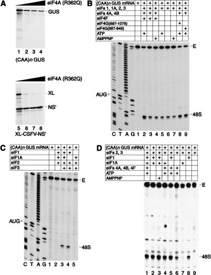
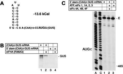
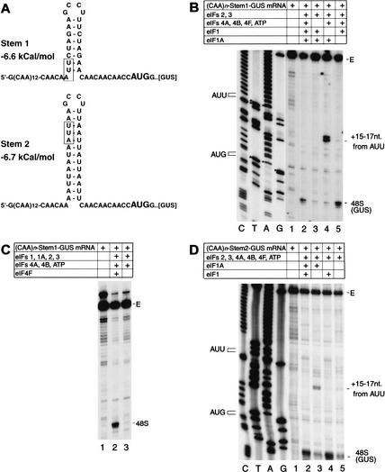
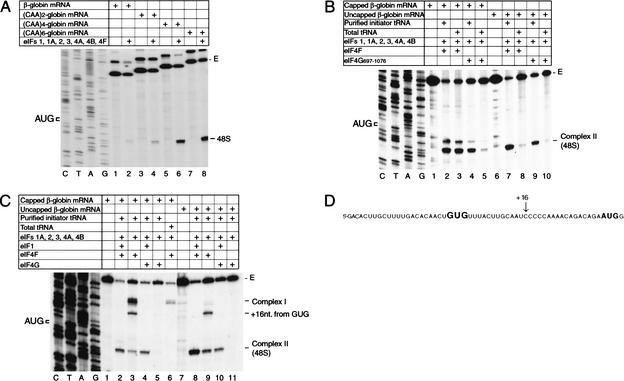
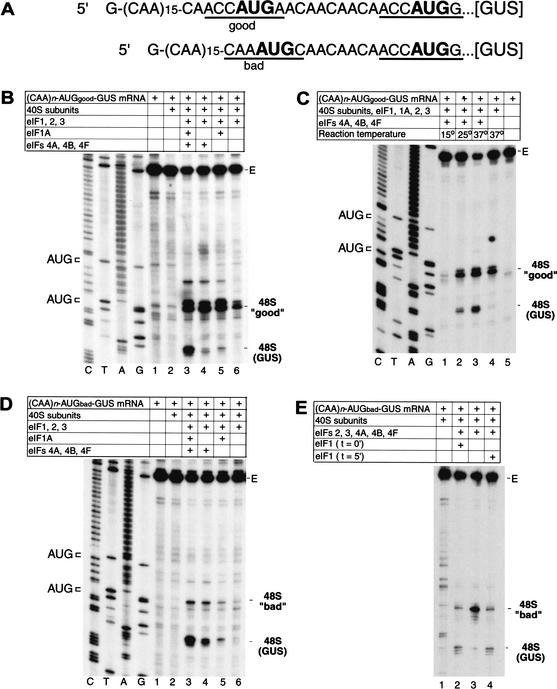
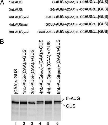
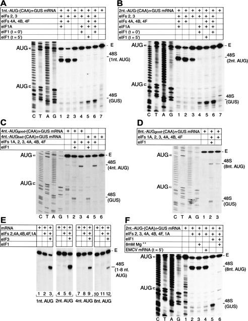
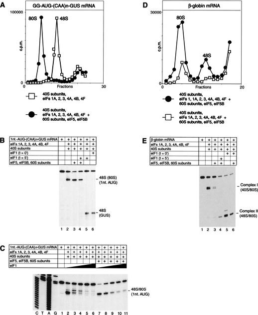
To elucidate an outline of the mechanism of eukaryotic translation initiation, 48S complex formation was analyzed on defined mRNAs in reactions reconstituted in vitro from fully purified translation components. We found that a ribosomal 40S subunit, eukaryotic initiation factor (eIF) 3, and the eIF2 ternary complex form a 43S complex that can bind to the 5'-end of an unstructured 5'-untranslated region (5'-UTR) and in the presence of eIF1 scan along it and locate the initiation codon without a requirement for adenosine triphosphate (ATP) or factors (eIF4A, eIF4B, eIF4F) associated with ATP hydrolysis. Scanning on unstructured 5'-UTRs was enhanced by ATP, eIFs 4A and 4B, and the central domain of the eIF4G subunit of eIF4F. Their omission increased the dependence of scanning on eIFs 1 and 1A. Ribosomal movement on 5'-UTRs containing even weak secondary structures required ATP and RNA helicases. eIF4F was essential for scanning, and eIFs 4A and 4B were insufficient to promote this process in the absence of eIF4F. We report that in addition to its function in scanning, eIF1 also plays a principal role in initiation codon selection. In the absence of eIF1, 43S complexes could no longer discriminate between cognate and noncognate initiation codons or sense the nucleotide context of initiation codons and were able to assemble 48S complexes on 5'-proximal AUG triplets located only 1, 2, and 4 nt from the 5'-end of mRNA.


Figures

Figure 1
Figure 1
Factor requirements for translation initiation on (CAA)n-GUS mRNA. (A) Partial resistance of (CAA)n-GUS mRNA translation to inhibition by R362Q mutant eukaryotic initiation factor (eIF) 4A. Uncapped (CAA)n-GUS (lanes 1–4) and XL-CSFV-NS‘ (lanes 5–8) mRNAs (0.2 μg) were translated in rabbit reticulocyte lysate (RRL; 15 μL) that had been preincubated with increasing amounts (0.3 μg, lanes 2,6; 1 μg, lanes 3,7; 2 μg, lanes 4,8) of mutant eIF4A at 30°C for 10 min, followed by addition of mRNA and further incubated at 30°C for 60 min. Lanes 1 and 5 did not contain added eIF4A. Samples were analyzed by SDS-PAGE and subsequent autoradiography. The positions of β-glucuronidase (GUS), XL, and NS‘ translation products are indicated to the right of appropriate panels. (B,C,D) Toeprint analysis of 48S complexes assembled on (CAA)n-GUS mRNA. Reaction mixtures contained 40S subunits, GMP-PNP, and either (B,C) aminoacylated total tRNA or (D) pure initiator tRNA in addition to translation components as indicated. Reaction mixture (C) did not contain adenosine triphosphate (ATP). Full-length cDNA is labeled E. cDNA products labeled “48S” terminated 15–17 nt downstream from the initiation codon of (CAA)n-GUS mRNA. The position of the initiation codon is shown to the left of the reference lanes, which show the (CAA)n-GUS sequence derived using the same primer as for toeprinting.
Figure 2
Figure 2
Translation initiation on 5′-stem-(CAA)n-GUS mRNA. (A) Secondary structure of the 5′-UTR of 5′-stem-(CAA)n-GUS mRNA as predicted using mfold version 3.0 (Mathews et al. 1999). The theoretical standard free energy of the secondary structure is indicated. (B) Translation of (CAA)n-GUS and 5′-stem-(CAA)n-GUS mRNAs (0.2 μg) in RRL (15 μL) that had been preincubated without added mutant eIF4A (lanes 1,3) or with 1 μg R362Q mutant eIF4A (lanes 2,4) under conditions as described in the legend to Figure 1A. (C) Toeprint analysis of 48S complex formation on 5′-stem-(CAA)n-GUS mRNA in reaction mixtures that contained 40S subunits, GMP-PNP, and aminoacylated total tRNA in addition to translation components as indicated. Full-length cDNA is labeled E. The label “48S” indicates the expected position of toeprints caused by assembled 48S complexes on this mRNA. The position of the initiation codon is shown to the left of the two reference lanes, which show 5′-stem-(CAA)n-GUS sequence derived using the same primer as for toeprinting.
Figure 3
Figure 3
Factor requirements for 48S complex formation on derivatives of (CAA)n-GUS mRNA containing AU-rich hairpins in the 5′-UTR. (A) Secondary structures of the 5′-UTRs of (CAA)n-Stem1-GUS and (CAA)n-Stem2-GUS mRNA as predicted using mfold version 3.0 showing the initiation codons (bold). The theoretical standard free energies of the secondary structures are indicated. AUU triplets in the 5′-UTR of these mRNAs are boxed. (B,C,D) Toeprinting analysis of dependence on initiation factors and ATP of 48S complex formation on (CAA)n-Stem1-GUS and (CAA)n-Stem2-GUS mRNAs. Reaction mixtures contained 40S subunits, GMP-PNP, and aminoacylated total tRNA in addition to translation components as indicated. Full-length cDNA is labeled E. cDNA products labeled “48S” terminated 15–17 nt downstream from the initiation codon of (CAA)n-Stem1-GUS or (CAA)n-Stem2-GUS mRNA, as appropriate. cDNA products labeled “+15–17 nt from AUU” terminated 15–17 nt downstream from indicated AUU triplets in the 5′-UTRs of these mRNAs. The position of AUU triplets and initiation codons are shown to the left of appropriate reference lanes, which show cDNA sequences derived using the same primers as for toeprinting.
Figure 4
Figure 4
48S complex formation on β-globin mRNA and β-globin mRNA derivatives. (A–C) Toeprinting analysis of 48S complex formation on natural 100% capped or in vitro transcribed uncapped β-globin mRNA and uncapped derivatives thereof containing progressively longer unstructured 5′-terminal sequences, as indicated, in reaction mixtures that contained 40S subunits, GMP-PNP, ATP, aminoacylated pure initiator tRNA or total tRNA, and translation components as indicated. The label “48S” indicates the position of toeprints caused by 48S complexes assembled on these mRNAs in panel A; the same complex is indicated as complex II (48S) in panels B and C. Complex I in panel C terminated 21–24 nt from the 5′ end; a complex in this panel that terminated 16 nt from a GUG triplet in the 5′-UTR shown in panel D is designated “+16 nt from GUG.” Full-length cDNA is labeled E, and the position of the initiation codon is shown to the left of the β-globin sequence in reference lanes in panels A–C. (D) Sequence of the 5′-UTR of β-globin mRNA showing the AUG initiation codon and a GUG triplet in the 5′-UTR in bold, as well as the position (+16) of the leading edge of 48S complexes assembled at the GUG triplet.
Figure 5
Figure 5
Factor dependence of initiation codon selection on mRNAs containing tandem initiation codons differing in sequence context. (A) Sequences of the 5′-UTRs of (CAA)n-AUGgood-GUS and (CAA)n-AUGbad-GUS mRNAs, showing the AUG initiation codons in bold. Context residues from −3 to +4 positions for each initiation codon are underlined; the A of the AUG codon is designated as +1. (B–E) Toeprinting analysis of 48S complex formation on (CAA)n-AUGgood-GUS and (CAA)n-AUGbad-GUS mRNAs, as indicated, in reaction mixtures containing 40S subunits, GMP-PNP, ATP, aminoacylated total tRNA, and translation components as indicated and incubated at 37°C (B,D,E) or at temperatures as indicated (C). eIF1 was included at the beginning of each assembly reaction except where indicated in panel E. Full-length cDNA is labeled E in panels B–E. The label “48S (GUS)” indicates the position of toeprints caused by 48S complexes assembled on the GUS initiation codon; the labels 48S “good” and 48S “bad” indicate 48 complexes assembled at upstream initiation codons in these mRNAs that are in either good or bad context. The position of these initiation codons are shown to the left of appropriate reference lanes (C,T,A,G) in panels B–D, which show cDNA sequences derived using the same primer as for toeprinting.
Figure 6
Figure 6
Initiation of translation from 5′-proximal initiation codons. (A) Sequences of the 5′-UTRs of 1nt-AUG-(CAA)n-GUS, 2nt-AUG-(CAA)n-GUS, 4nt-AUGgood-(CAA)n-GUS, 4nt-AUGbad-(CAA)n-GUS, and 8nt-AUGgood-(CAA)n-GUS mRNAs, showing the AUG initiation codons in bold. (B) Products of translation in RRL of these mRNAs and of (CAA)n-GUS mRNA, as indicated. Translation was performed under standard conditions. 5′AUG and GUS indicate the products of translation that were initiated at upstream 5′-proximal initiation codons and at the GUS initiation codon, respectively.
Figure 7
Figure 7
Factor dependence of initiation codon selection on derivatives of (CAA)n-GUS mRNA containing 5′-proximal initiation codons. (A–F) Toeprinting analysis of 48S complex formation on mRNAs containing 5′-proximal initiation codons, as indicated, in reaction mixtures containing 40S subunits, GMP-PNP, ATP, aminoacylated total tRNA, and translation components as indicated. eIF1 was included at the beginning of each assembly reaction except where indicated in panels A and B. Encephalomyocarditis virus (EMCV) competitor mRNA (5 μg) was added to a reaction after 5 min of incubation where indicated on panel F. MgAc (8 mM) was added to reactions where indicated on panel F. Full-length cDNA is labeled E. The label “48S GUS” indicates the position of toeprints caused by 48S complexes assembled on the GUS initiation codon; other labels such as 48S (1 nt AUG) indicate 48 complexes assembled at the indicated 5′-proximal initiation codon in these mRNAs. The positions of initiation codons are shown to the left of appropriate reference lanes (C,T,A,G) in panels A–D and F, which also show cDNA sequences derived using the same primer as for toeprinting.
Figure 8
Figure 8
Differences between 48S complexes assembled on cap-proximal initiation codons and “complex I” aberrantly assembled on natural capped globin mRNA in subunit joining activities and in protection by 60S subunits from eIF1-induced destabilization. (A,D) Sucrose density gradient centrifugation of 48S and 80S complexes assembled on [32P]-labeled (A) GG-AUG-(CAA)n-GUS mRNA and (D) globin mRNA from purified translation components as indicated. Sedimentation was from right to left. (B,E) Effect of inclusion of eIF1 at indicated times to assembly reactions that contained (B) G-AUG-(CAA)n-GUS mRNA and (E) β-globin mRNA, GTP, aminoacylated tRNA, and other translation components as indicated. (C) Effect of inclusion of increasing amounts (0.05 μg, lanes 3,8; 0.15 μg, lanes 4,9; 0.45 μg, lanes 5,10; 1 μg, lanes 6,11) of eIF1 in assembly reactions that contained G-AUG-(CAA)n-GUS mRNA, GTP, aminoacylated tRNA, and other translation components as indicated. Lanes 2 and 7 did not contain added eIF1.

Similar articles

Cited by

References

    1. Ali IK, McKendrick L, Morley SJ, Jackson RJ. Truncated initiation factor eIF4G lacking an eIF4E binding site can support capped mRNA translation. EMBO J. 2001;20:4231–4244. - PMC - PubMed
    1. Anthony DD, Merrick WC. Analysis of 40S and 80S complexes with mRNA as measured by sucrose density gradients and primer extension inhibition. J Biol Chem. 1992;267:1554–1562. - PubMed
    1. Banerjee AK. 5′-terminal cap structure in eucaryotic messenger ribonucleic acids. Microbiol Rev. 1980;44:175–205. - PMC - PubMed
    1. Battiste JL, Pestova TV, Hellen CUT, Wagner G. The eIF1A solution structure reveals a large RNA-binding surface important for scanning function. Mol Cell. 2000;5:109–119. - PubMed
    1. Carberry SE, Goss DJ. Interaction of wheat germ protein synthesis initiation factors eIF-3, eIF-(iso)4F, and eIF-4F with mRNA analogues. Biochemistry. 1991;30:6977–6982. - PubMed

Publication types

MeSH terms