. 2002 Nov 15;16(22):2958-70.
doi: 10.1101/gad.240102.

Nephric lineage specification by Pax2 and Pax8

Affiliations

Nephric lineage specification by Pax2 and Pax8

Maxime Bouchard et al. Genes Dev. .

Abstract

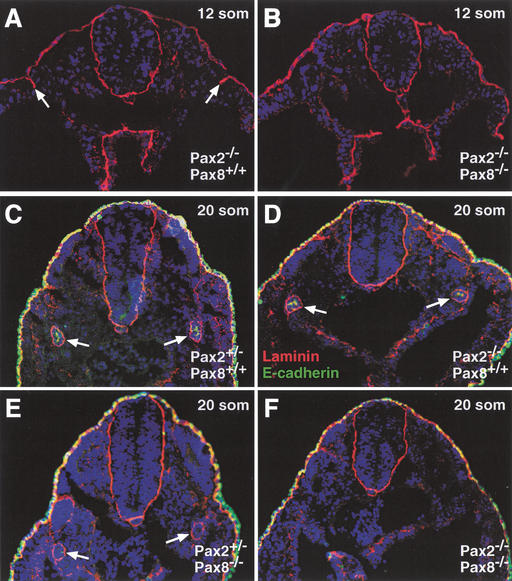
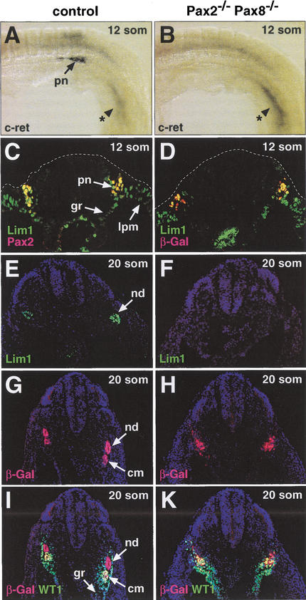
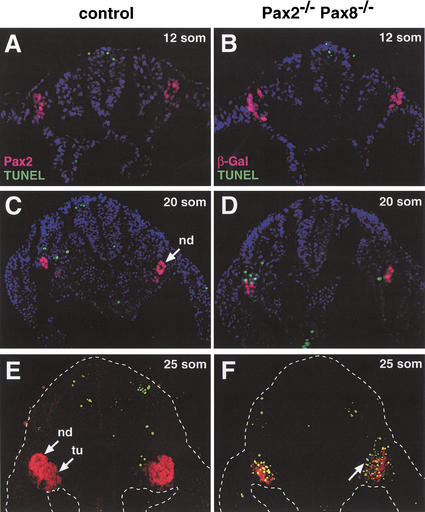
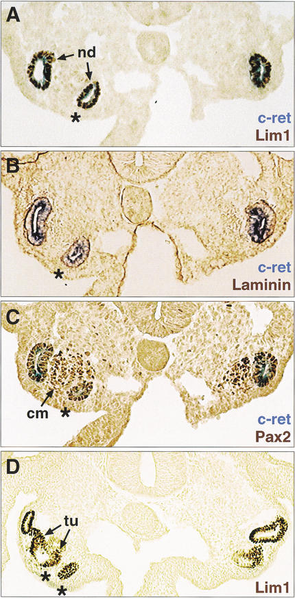
The mammalian kidney develops in three successive steps from the initial pronephros via the mesonephros to the adult metanephros. Although the nephric lineage is specified during pronephros induction, no single regulator, including the transcription factor Pax2 or Pax8, has yet been identified to control this initial phase of kidney development. In this paper, we demonstrate that mouse embryos lacking both Pax2 and Pax8 are unable to form the pronephros or any later nephric structures. In these double-mutant embryos, the intermediate mesoderm does not undergo the mesenchymal-epithelial transitions required for nephric duct formation, fails to initiate the kidney-specific expression of Lim1 and c-Ret, and is lost by apoptosis 1 d after failed pronephric induction. Conversely, retroviral misexpression of Pax2 was sufficient to induce ectopic nephric structures in the intermediate mesoderm and genital ridge of chick embryos. Together, these data identify Pax2 and Pax8 as critical regulators that specify the nephric lineage.


Figures

Figure 1
Figure 1
Inactivation of the Pax8 locus by insertion of a cre gene. (A) Structure of the wild-type and targeted Pax8 loci. The cre gene was fused in frame to Pax8 exon 3 followed by a neomycin (neo) resistance gene and an SV40 polyadenylation site (pA). The herpes simplex virus thymidine kinase (tk) and diphteria toxin A (DT-A) genes were used for counterselection against random integration in ES cells. The neo expression cassette was flanked by frt sites (red arrowheads) that mediate deletion by the FLP recombinase. Correct targeting was verified by Southern blot analysis of EcoRI-digested DNA with the indicated probe. The lengths of DNA fragments are indicated in kilobases. The Pax8 exons are numbered according to Okladnova et al. (1997) with exon 3 coding for the N-terminal part of the paired domain. The Pax8neo allele codes for a fusion protein consisting of the first 12 amino acids of Pax8 (underlined) linked to the SV40 nuclear localization signal and the N-terminal Cre protein sequences (boldface type): MPHNSIRSGHGGPKKKRKVSNLL. E, EcoRI; N, NcoI; Sa, SacI; Ss, SspI. (B) Southern blot analysis of EcoRI-digested tail DNA from wild-type (+/+) and heterozygous (+/−) Pax8 mutant mice.
Figure 2
Figure 2
Development of the urogenital system in Pax2,Pax8 mutant embryos. (AD) The urogenital system of male E18.5 embryos of the indicated genotypes was dissected and photographed. The hypoplastic kidneys of Pax2+/−Pax8+/− embryos (C) were approximately fourfold smaller than control kidneys (A,B). Pax2+/−Pax8−/− embryos (D) failed to develop a kidney, ureter, and genital tract (vas deferens), whereas the adrenal gland, testis, and bladder formed normally. (E,F) Kidney sections stained with hematoxylin and eosin. The nephric tubules and glomeruli were reduced in number and the stromal component was increased in the hypoplastic Pax2+/−Pax8+/− kidney (F) compared with the control embryo (E) at E18.5. a, adrenal gland; b, bladder; g, glomerulus; k, kidney; s, S-shaped body; st, stroma; t, testis; tu, tubule; u, ureter; ut, ureteric tip; v, vas deferens.
Figure 3
Figure 3
Pax2-independent expression of Pax8 at the onset of kidney development. (A,B) Initiation of Pax gene expression in the pronephric anlage of Pax2+/− embryos. As shown by whole-mount in situ hybridization, Pax8 expression (B) was first detected at the 7-somite stage in the intermediate mesoderm at the level of the fifth and sixth somites corresponding to the pronephric anlage. At the 9-somite stage, the Pax2 gene (A) was also expressed in the pronephric anlage, where its expression was first observed at 8 somites (data not shown). (C) Coexpression of Pax2 and Pax8 in the pronephric anlage. Pax2 protein (brown) and Pax8 transcripts (blue) were simultaneously detected on a transverse section of a 10-somite Pax2+/− embryo by immunostaining and in situ hybridization, respectively. (D) Absence of epithelial cells in the pronephric anlage at 10 somites. An adjacent section of the same embryo shown in (C) was stained with an anti-laminin antibody (brown) in combination with Pax8 in situ hybridization (blue). (E,F) Pax2-independent initiation and maintenance of Pax8 expression during kidney development. Pax8 transcripts were detected by whole-mount in situ hybridization of Pax2+/− and Pax2−/− embryos at 9 (E) and 19 (F) somites. ba, branchial arch; mhb, midbrain-hindbrain boundary; ms, mesonephros; nt, neural tube; op, otic placode; ov, otic vesicle; pa, pronephric anlage; som, somite.
Figure 4
Figure 4
Early defects of pro- and mesonephros development in Pax2−/−Pax8−/− embryos. (AD) The expression of β-galactosidase from the mutant Pax2 allele (Bouchard et al. 2000) was used as a kidney-specific marker to visualize pro- and mesonephros development in Pax2,Pax8 mutant embryos of the indicated genotypes. Embryos of the same litter, which developed to the 14- (C,D) or 15- (A,B) somite stage, were stained with X-gal for the same period of time (15 h). Pax2+/− (A) and Pax2−/− (B) embryos expressed β-galactosidase activity in the pronephros (pn) and mesonephros (ms) extending from somite 9 to just beyond the last somite, 15. The β-galactosidase expression in Pax2−/−Pax8+/− embryos (C) was restricted to a region between somites 9 and 13. The absence of any functional Pax2/8 allele led to the down-regulation of β-galactosidase expression (arrowheads) in Pax2−/−Pax8−/− embryos (D). It is important to note that the presence of a single Pax2 (lacZ) allele resulted in a disproportionately lower X-gal staining signal in Pax2+/− embryos (A) compared with Pax2−/− embryos (B,C), which expressed the lacZ gene only at a twofold higher level because of the presence of two lacZ alleles. This nonlinearity of the β-galactosidase assay is also the reason why the reduced lacZ expression in Pax2−/−Pax8−/− embryos (D) generated a low X-gal staining signal, although it could be readily detected by the more sensitive, but nonquantitative antibody staining method (Fig. 6H).
Figure 5
Figure 5
Absence of mesenchymal-epithelial transitions in the intermediate mesoderm of Pax2−/−Pax8−/− embryos. The expression of the epithelial markers laminin (red) and E-cadherin (green) was analyzed in embryos of the indicated genotypes at 12 (A,B) or 20 (CF) somites by immunostaining of transverse sections. At 12 somites, epithelial cells were bilaterally present in the pronephric region of Pax2−/−Pax8+/+ embryos (arrows in A) in contrast with Pax2−/−Pax8−/− embryos (B). At the 20-somite stage, a distinct ring of laminin expression demarcates the nephric duct of all mutant embryos (arrows in CE) except in Pax2−/−Pax8−/− embryos (F). E-cadherin expression was reduced in the nephric duct of Pax2+/−Pax8−/− embryos (E; additional data not shown) and absent in Pax2−/−Pax8−/− embryos (F).
Figure 6
Figure 6
Absence of Lim1 and c-Ret expression in the intermediate mesoderm of Pax2−/−Pax8−/− embryos. (AD) c-Ret and Lim1 expression at the 12-somite stage. c-Ret transcripts were detected by whole-mount in situ hybridization in the pronephros of wild-type control embryos (A) in contrast to Pax2−/−Pax8−/− embryos (B) that, however, still expressed c-ret in the tail region (indicated by asterisk). Antibody staining of transverse sections was used to reveal the expression of Lim1 and Pax2 or β-galactosidase (β-Gal) in control Pax8+/− (C) and Pax2−/−Pax8−/− (D) embryos, respectively. (EK) Lim1 and WT1 expression at the 20-somite stage. Adjacent transverse sections of control Pax2+/−and Pax2−/−Pax8−/− embryos were analyzed by immunostaining for Lim1 (E,F), β-Gal (GK) and WT1 (I,K) protein expression. cm, condensing mesenchyme; gr, genital ridge; lpm, lateral plate mesoderm; nd, nephric duct; pn, pronephros.
Figure 7
Figure 7
Late apoptosis of intermediate mesoderm cells in Pax2−/−Pax8−/− embryos. Cells undergoing apoptosis (green dots) were detected on transverse sections of control or Pax2−/−Pax8−/− embryos by TUNEL assay in combination with immunohistochemical analysis of Pax2 (red; A,C,E) or β-galactosidase (red; B,D,F) expression. Apoptotic cells within or adjacent to the β-Gal (Pax2) expression domain were absent in Pax2−/−Pax8−/− embryos at 12 somites (B), slightly increased at 20 somites (D) and abundant at 25 somites (F) compared with the control Pax2+/−(C) and Pax8+/− (A,E) embryos. An arrow in F points to apoptotic cells surrounding the β-Gal expression domain. nd, nephric duct; tu, tubules.
Figure 8
Figure 8
Ectopic kidney formation on Pax2 misexpression in the intermediate mesoderm. A chicken retrovirus expressing the mouse Pax2 protein (RCAS–mPax2) was injected into the mid-streak tissue adjacent to Hensen's node on one side of chick embryos at HH stage 4–6. (AC) Ectopic kidney formation in a representative embryo at 48 h after injection (HH stage 17–18). Adjacent sections were stained by in situ hybridization for c-ret mRNA expression (blue; AC) and by immunohistochemistry (brown) for Lim1 (A), laminin (B) or Pax2 (C) protein expression. (D) Lim1 staining of an embryo that progressed further in kidney development. Asterisks denote ectopic nephric ducts and tubules that formed in the region of the genital ridge on the injected side (left). cm, condensing mesenchyme; nd, nephric duct; tu, tubules.

References

    1. Adams B, Dörfler P, Aguzzi A, Kozmik Z, Urbánek P, Maurer-Fogy I, Busslinger M. Pax-5 encodes the transcription factor BSAP and is expressed in B lymphocytes, the developing CNS, and adult testis. Genes Dev. 1992;6:1589–1607. - PubMed
    1. Barnes JD, Crosby JL, Jones CM, Wright CVE, Hogan BLM. Embryonic expression of Lim-1, the mouse homolog of Xenopus XLim-1, suggests a role in lateral mesoderm differentiation and neurogenesis. Dev Biol. 1994;161:168–178. - PubMed
    1. Bouchard M, Pfeffer P, Busslinger M. Functional equivalence of the transcription factors Pax2 and Pax5 in mouse development. Development. 2000;127:3703–3713. - PubMed
    1. Bouchard M, Schleiffer A, Eisenhaber F, Busslinger M. Evolution and function of Pax genes. In: Cooper D, editor. Encyclopedia of the Human Genome. UK: Nature Publishing Group; 2003. . (In Press.)
    1. Brophy PD, Ostrom L, Lang KM, Dressler GR. Regulation of ureteric bud outgrowth by Pax2-dependent activation of the glial derived neurotrophic factor gene. Development. 2001;128:4747–4756. - PubMed

Publication types

MeSH terms

Associated data