

doi: 10.1186/1476-5926-1-1.
Structural and functional aspects of liver sinusoidal endothelial cell fenestrae: a review
Affiliations
- PMID: 12437787
- PMCID: PMC131011
- DOI: 10.1186/1476-5926-1-1
Item in Clipboard
Structural and functional aspects of liver sinusoidal endothelial cell fenestrae: a review
Comp Hepatol.
.
Abstract
This review provides a detailed overview of the current state of knowledge about the ultrastructure and dynamics of liver sinusoidal endothelial fenestrae. Various aspects of liver sinusoidal endothelial fenestrae regarding their structure, origin, species specificity, dynamics and formation will be explored. In addition, the role of liver sinusoidal endothelial fenestrae in relation to lipoprotein metabolism, fibrosis and cancer will be approached.
Figures
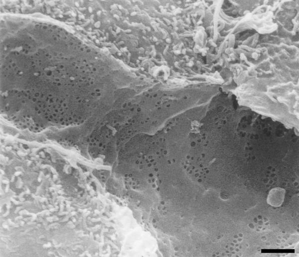
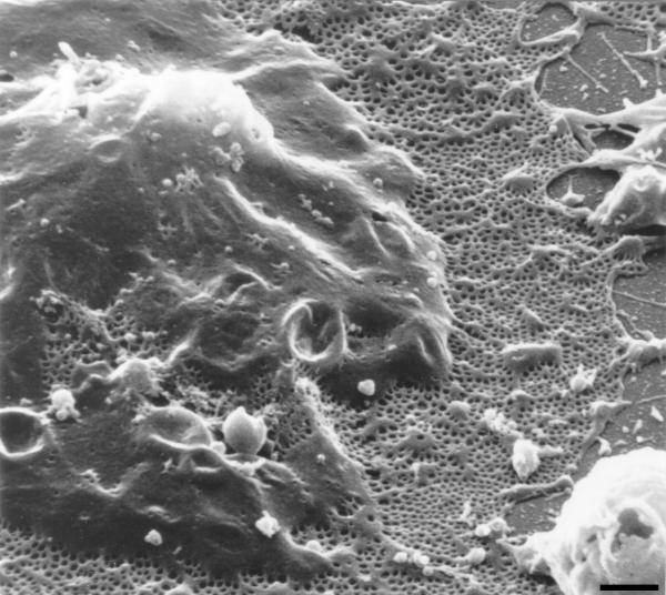
Low magnification scanning electron micrograph of the sinusoidal endothelium from rat liver showing the fenestrated wall. Notice the clustering of fenestrae in sieve plates. Scale bar, 1 μm.
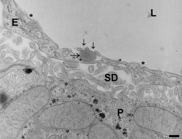
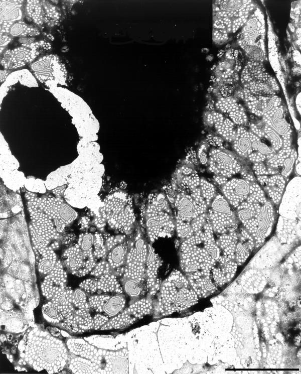
High-magnification transmission electron micrograph of a hepatic sinusoid of rat liver, fixed by perfusion-fixation with glutaraldehyde, postfixed in osmium, dehydrated in alcohol, and embedded in Epon (reference[1]). The lumen of the sinusoid (L) is lined by the endothelium (E), showing the presence of fenestrae (small arrows) and coated pits (asterisks). Note a lipid particle (large arrow) which passed the fenestrae, illustrating the sieving effect of fenestrae. The space of Disse (SD) contains numerous microvilli of the parenchymal cells (P). (Courtesy of Drs R. De Zanger, reference [7]). Scale bar, 300 nm.
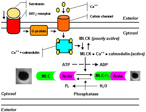
Scheme of the serotonin signal pathway showing the steps in fenestral contraction and relaxation, as postulated by Gatmaitan et al.[89,90,104]: Serotonin binds to a ketanserin-inhibitable receptor, coupled to a pertussis-toxin sensitive G-protein; a calcium channel opens, causing an influx of calcium ions; the intracellular calcium level increases rapidly, and calcium binds to calmodulin; the calcium-calmodulin complex activates myosin light chain kinase, and as a result phosphorylation of the 20-kd light chain of myosin occurs, resulting in an increased actin-activated myosin ATPase activity, which finally initiates contraction of fenestrae. The mechanism for the relaxation of LSEC fenestrae is presently unclear and probably involves dephosphorylation of myosin light chains as represented by dashed lines: a decrease in the cytosolic free calcium concentration leads to dissociation of calcium and calmodulin from the kinase, thereby inactivating myosin light chain kinase, under these conditions myosin light chain phosphatase, which is not dependent on calcium for activity, dephosphorylates myosin light chain and finally causes relaxation of fenestrae.
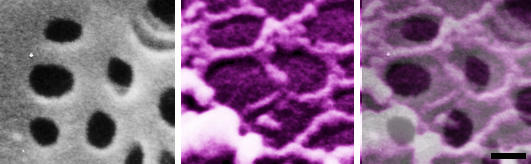
High-power scanning electron micrographs of a nonextracted (left); and of a formaldehyde prefixed, cytoskeleton buffer extrated rat liver sinusoidal endothelial cell (middle). Notice the grouped fenestrae on the cell surface (left); and a remarkable series of rings of fenestrae-associated cytoskeleton (middle). Layering a colored scanning electron micrograph on top of the cell surface of a nonextracted rat liver sinusoidal endothelial cell clearly illustrates the relation between both structures (right). Scale bar, 200 nm.
Scanning electron micrograph of an LSEC treated with 0.1 μg/ml of latrunculin A for 2 hours, showing huge fenestrated areas. Thin cytoplasmic arms divide flat fields containing numerous fenestrae. The bulging area corresponds with the nucleus. Scale bar, 2 μm.
Whole-mount transmission electron micrograph of an LSEC treated with 100 nM dihydrohalichondramide for 1 hour, showing the dark nuclear area and surrounding extracted cytoplasm. Note the presence of small cytoplasmatic areas of intermediated density within the fenestrated cytoplasm. In several of these areas a very peculiar structure could be observed, consisting of rows of fenestrae with increasing diameter, fanning out into the surrounding cytoplasm, connected to the small cytoplasmatic areas with their smallest fenestrae. These structures are suggestive of de novo fenestrae formation and we therefore named them "fenestrae-forming center" (FFC) [79]. Scale bar, 5 μm.
References
-
- Wisse E. An electron microscopic study of the fenestrated endothelial lining of rat liver sinusoids. J Ultrastruct Res. 1970;31:125–150. - PubMed
-
- Ogawa K, Minase T, Enomoto K, Onoé T. Ultrastructure of fenestrated cells in the sinusoidal wall of rat liver after perfusion fixation. Tohoku J Exp Med. 1973;110:89–101. - PubMed
-
- Wisse E, De Zanger RB, Charels K, Van Der Smissen P, McCuskey RS. The liver sieve: Considerations concerning the structure and function of endothelial fenestrae, the sinusoidal wall and the space of Disse. Hepatology. 1985;5:683–692. - PubMed
-
- Wisse E, De Zanger RB, Jacobs R, McCuskey RS. Scanning electron microscope observations on the structure of portal veins, sinusoids and central veins in rat liver. Scan Electron Microsc. 1983;3:1441–1452. - PubMed
LinkOut - more resources
Full Text Sources
Other Literature Sources