Construction of a novel transposon mutagenesis system useful in the isolation of Streptococcus parasanguis mutants defective in Fap1 glycosylation
- PMID: 12438322
- PMCID: PMC132963
- DOI: 10.1128/IAI.70.12.6534-6540.2002
Construction of a novel transposon mutagenesis system useful in the isolation of Streptococcus parasanguis mutants defective in Fap1 glycosylation
Abstract
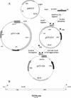
Streptococcus parasanguis, a primary colonizer of the tooth surface, has long, peritrichous fimbriae. A fimbria-associated protein, Fap1, is identified as an adhesin of S. parasanguis FW213. The mature Fap1 protein is glycosylated, and the glycosylation is required for fimbria biogenesis and bacterial adhesion. Little is known about the mechanism of Fap1 glycosylation due to the lack of identifiable mutants. A novel transposon mutagenesis system was established and used to generate a mutant library. Screening of the library with a monoclonal antibody specific for a glycan epitope of Fap1 yielded six mutants with decreased expression levels of surface-associated glycosylated Fap1 protein. Southern blot analyses revealed that three of the mutants had the transposon inserted in the fap1 locus, whereas the other three mutants had insertions in other genes. Among the latter three mutants, two expressed Fap1 polypeptides on which no glycosylation was detected by glycan-specific antibodies; the other mutant expressed a partially glycosylated Fap1 polypeptide. These data suggest that three mutants were isolated with defects in genes implicated in Fap1 glycosylation.
Figures


References
-
- Benz, I., and M. A. Schmidt. 2001. Glycosylation with heptose residues mediated by the aah gene product is essential for adherence of the AIDA-I adhesin. Mol. Microbiol. 40:1403-1413. - PubMed
-
- Boyer, H. W., and D. Roulland-Dussoix. 1969. A complementation analysis of the restriction and modification of DNA in Escherichia coli. J. Mol. Biol. 41:459-472. - PubMed
-
- Byrne, M. E., D. A. Rouch, and R. A. Skurray. 1989. Nucleotide sequence analysis of IS256 from the Staphylococcus aureus gentamicin-tobramycin-kanamycin-resistance transposon Tn4001. Gene 81:361-367. - PubMed
-
- Calabi, E., S. Ward, B. Wren, T. Paxton, M. Panico, H. Morris, A. Dell, G. Dougan, and N. Fairweather. 2001. Molecular characterization of the surface layer proteins from Clostridium difficile. Mol. Microbiol. 40:1187-1199. - PubMed
Publication types
MeSH terms
Substances
Grants and funding
LinkOut - more resources
Full Text Sources

