. 2002 Dec;70(12):6597-605.
doi: 10.1128/IAI.70.12.6597-6605.2002.

Leishmania pifanoi pathogenesis: selective lack of a local cutaneous response in the absence of circulating antibody

Affiliations

Leishmania pifanoi pathogenesis: selective lack of a local cutaneous response in the absence of circulating antibody

María Colmenares et al. Infect Immun. 2002 Dec.

Abstract

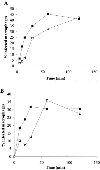
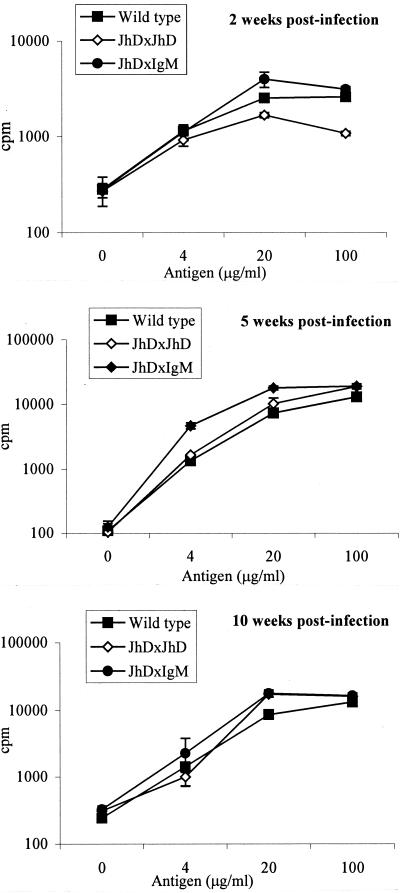
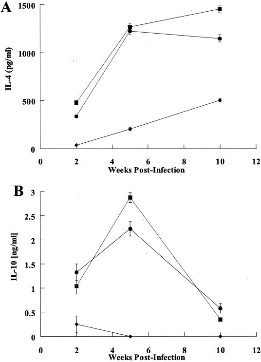
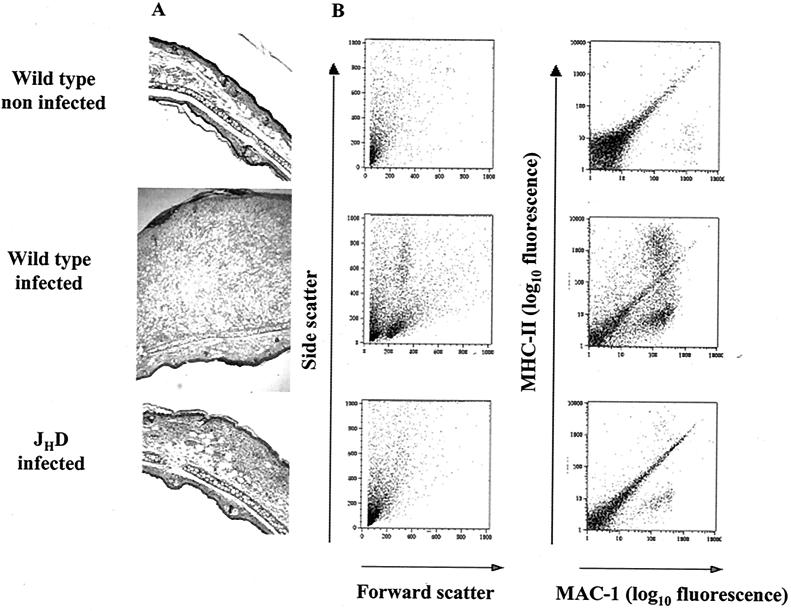
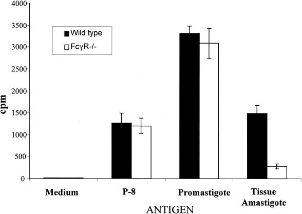
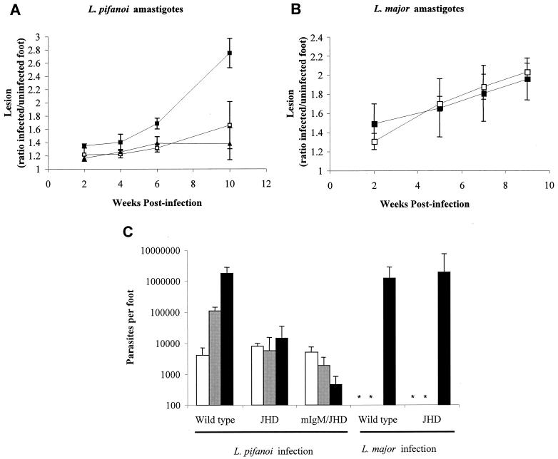
Recently, a role for B cells in the pathogenesis associated with infection by Leishmania (Leishmania mexicana complex and L. donovani) has been established. In the case of L. mexicana complex parasites (L. mexicana, L. pifanoi, and L. amazonensis), a critical role for immunoglobulin G-mediated mechanisms for the amastigote stage in the host is evident; however, the immunological mechanisms involved remain to be established. In vitro analysis of the kinetics of parasite uptake by macrophages failed to indicate a major effect of antibody opsonization. Given the importance of CD4(+) T cells in the development of disease caused by these parasites, the possibility that the lack of pathogenesis was due to the lack of development of an immune response at the local site (draining lymph node and/or cutaneous site) was explored. Interestingly, the level of CD4(+)-T-cell activation (proliferation and cytokine) in draining lymph nodes from mice lacking circulating antibody (resistant) was found to be comparable to that in nodes from wild-type mice (susceptible) at 2, 5, and 10 weeks postinfection. However, antibody-deficient animals had markedly reduced numbers of monocytes and lymphocytes recruited or retained at the site of cutaneous infection in comparison to wild-type mice, indicating a selective impairment in the local cutaneous immune response. In vitro antigen presentation studies employing tissue-derived (opsonized) amastigotes demonstrated that L. pifanoi-infected FcR(-/-) macrophages, in contrast to comparably infected wild-type cells, failed to activate Leishmania antigen-specific T lymphocytes. These data, taken together, suggest that one possible mechanism for the role of antibody in pathogenesis may be to mediate parasite uptake and regulate the immune response at the local cutaneous site of infection.


Figures

FIG. 1.
FIG. 1.
Kinetics of macrophage infection with L. pifanoi. (A) J774A.1 macrophages were incubated with L. pifanoi axenic amastigote (□) or amastigotes opsonized with antibodies (▪). (B) Peritoneal resident macrophages from wild-type BALB/c (▪) or BALB/c FcγR−/− mice (□) were incubated with tissue-derived amastigote. Infections were carried out at a ratio of 1:1 of parasites to macrophages. At indicated time points, cells were fixed and stained with DAPI, and the percentage of infected macrophages was determined microscopically from the analysis of 200 phagocytic cells. These results are representative of three independent determinations.
FIG. 2.
FIG. 2.
Recall responses of CD4+ T cells obtained from lymph nodes of L. pifanoi amastigote-infected mice. CD4+ T cells were recovered from infected wild-type BALB/c, JHD, or mIgM/JHD mice at 2, 5, and 10 weeks postinfection. The cells were then stimulated in an in vitro proliferation experiment as described in Materials and Methods using T-cell-depleted splenocytes as APCs. Bars represent standard deviation. These results are representative of two separate experiments.
FIG. 3.
FIG. 3.
Cytokine production by proliferating CD4+ T cells. CD4+ T cells were recovered from infected wild-type BALB/c (▪), JHD (♦), or mIgM/JHD (•) mice at different times postinfection. The cells were then stimulated in an in vitro proliferation experiment (100 μg of antigen) as described in Materials and Methods, and the supernatants were analyzed for IL-4 (A) or IL-10 (B) production by enzyme-linked immunosorbent assay. It should be noted that, in all cases, the levels of secreted IFN-γ were below detection (data not shown). These results are representative of two separate determinations. Bars represent standard deviation.
FIG. 4.
FIG. 4.
Analysis of leukocyte recruitment in the cutaneous lesion site. Mice were infected intradermally with 2 × 10⁶ L. pifanoi axenic amastigotes, and at different times postinfection, ears were recovered. (A) Hematoxylin- and eosin-stained transverse sections; at 5 weeks postinfection, serial sections through the inoculation site were prepared, and the section showing the greatest transverse thickness was chosen for presentation. Magnification, ×100. (B) Representation of two-parameter dot plots of epidermal cells recovered from mice at 10 weeks postinfection and prepared for flow cytometry analysis as described in Materials and Methods. Cells were identified as follows: lymphocytes, medium size (forward scatter) and medium granular (side scatter); monocytes, positive for MAC-1 and negative or low for MHC-II; dendritic leukocytes and activated macrophages (APCs), positive for MAC-1 and for MHC-II. The quantification for individual cell populations (CD4+ and/or CD8+ T cells or macrophages) is shown in Table 1. These results are representative of three independent determinations.
FIG. 5.
FIG. 5.
Antigen presentation of Leishmania antigen to CD4+ T cells by infected macrophages from BALB/c FcγR−/− or from wild-type BALB/c mice. The proliferative responses of CD4+ T cells from mice sensitized with frozen and thawed L. pifanoi amastigote antigen were employed to assess antigen presentation. PECs from FcγR−/− mice (□) or from BALB/c (▪) mice were infected with tissue-derived amastigotes at a ratio of 5:1 of parasites to macrophages, and their capacity to present endogenously synthesized Leishmania antigen to CD4+ T cells was assessed after 16 h of infection. At 16 h postinfection, the level of infected macrophages (as assessed by DAPI staining) was comparable for BALB/c-FcγR−/− and wild-type macrophages. The presentation of an affinity-purified Leishmania antigen, P-8 (1 μg/ml), and parasite antigen presentation after infection with promastigotes were used as controls in these experiments. Bars represent standard deviations. These results are representative of two independent experiments.
FIG. 6.
FIG. 6.
Course of infection by either L. pifanoi (A) or L. major (B) amastigotes in B-cell-deficient mice. The course of infection in JHD (□), mIgM/JHD (▴), or wild-type BALB/c (▪) mice was monitored after infection by measuring lesion development. Each data point is the mean from six to eight mice, and the bars represent standard errors of the means. (C) Parasite burdens from footpads of infected mice. At 2 (□), 5 (▪), and 10 (▪) weeks postinfection, infected feet from mice inoculated with L. pifanoi or L. major amastigotes were quantitatively assessed for parasites by a limiting dilution assay. Each data point is the mean from three mice, and the bars represent standard errors of the means. *, not determined. These results are representative of two separate experiments.

References

    1. Afonso, L. C., and P. Scott. 1993. Immune responses associated with susceptibility of C57BL/10 mice to Leishmania amazonensis. Infect. Immun. 61:2952-2959. - PMC - PubMed
    1. Alonso, A., Y. Bayon, M. Renedo, and M. S. Crespo. 2000. Stimulation of Fc gamma R receptors induces monocyte chemoattractant protein-1 in the human monocytic cell line THP-1 by a mechanism involving I kappa B-alpha degradation and formation of p50/p65 NF-kappa B/Rel complexes. Int. Immunol. 12:547-554. - PubMed
    1. Balestieri, F. M., A. R. Queiroz, C. Scavone, V. M. Costa, M. Barral-Netto, and A. Abrahamsohn Ide. 2002. Leishmania (L.) amazonensis-induced inhibition of nitric oxide synthesis in host macrophages. Microbes Infect. 4:23-29. - PubMed
    1. Barral-Netto, M., A. Barral, C. E. Brownell, Y. A. Skeiky, L. R. Ellingsworth, D. R. Twardzik, and S. G. Reed. 1992. Transforming growth factor-beta in leishmanial infection: a parasite escape mechanism. Science 257:545-548. - PubMed
    1. Belkaid, Y., K. F. Hoffmann, S. Mendez, S. Kamhawi, M. C. Udey, T. A. Wynn, and D. L. Sacks. 2001. The role of interleukin (IL)-10 in the persistence of Leishmania major in the skin after healing and the therapeutic potential of anti-IL-10 receptor antibody for sterile cure. J. Exp. Med. 194:1497-1506. - PMC - PubMed

Publication types

Substances