. 2002 Dec;70(12):6646-51.
doi: 10.1128/IAI.70.12.6646-6651.2002.

EndoS and SpeB from Streptococcus pyogenes inhibit immunoglobulin-mediated opsonophagocytosis

Affiliations

EndoS and SpeB from Streptococcus pyogenes inhibit immunoglobulin-mediated opsonophagocytosis

Mattias Collin et al. Infect Immun. 2002 Dec.

Abstract

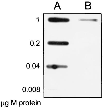
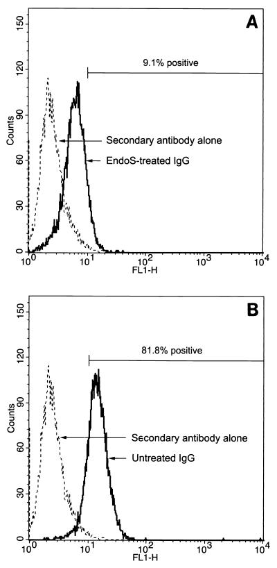
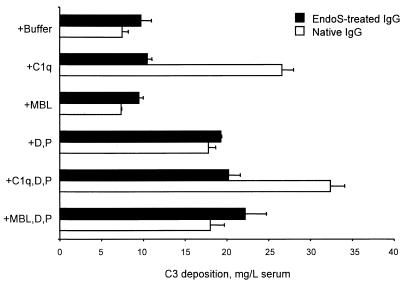
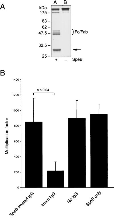
The human pathogen Streptococcus pyogenes primarily infects the upper respiratory tract and skin, but occasionally it disseminates and causes severe invasive disease with high mortality. This study revealed that the activity of extracellular EndoS, which hydrolyzes the functionally important N-linked oligosaccharides on opsonizing immunoglobulin G (IgG), contributes to increased survival of S. pyogenes in human blood ex vivo. The inability to kill the bacteria is due to reduced binding of IgG to Fc receptors and impaired classical pathway-mediated activation of complement. In addition, the activity of extracellular SpeB, which cleaves IgG into Fc and Fab fragments, also increases bacterial survival. This suggests that S. pyogenes expresses two enzymes, EndoS and SpeB, which modulate IgG by different mechanisms in order to evade the adaptive immune system.


Figures

FIG. 1.
FIG. 1.
Purified IgGops binds to purified M1 protein. Serial dilutions of M1 protein were applied to a membrane in a slot-binding experiment, followed by incubation with IgGops (lane A) or nonopsonizing IgG (lane B). IgG was subsequently detected with peroxidase-labeled protein A.
FIG. 2.
FIG. 2.
rEndoS hydrolysis of IgGops promotes bacterial growth in human blood. (A) SDS-PAGE and lectin blot analysis of IgG incubated with (+) or without (−) rEndoS. Lanes A and C, IgG incubated with rEndoS; lanes B and D, IgG incubated with PBS alone. (B) Representative results of opsonophagocytic killing assays performed with rEndoS-treated IgG from a donor. IgG treated with rEndoS (rEndoS-treated IgG) or IgG treated with PBS (intact IgG) was added to human blood. Alternatively, no IgG or rEndoS alone was added. Bacterial growth was monitored after 3 h of incubation. The mean multiplication factor (n = 5) and standard deviations are shown.
FIG. 3.
FIG. 3.
rEndoS-treated IgG binds poorly to leukocytes. Binding of rEndoS-treated (A) and intact IgG (B) to U937 cells was analyzed by flow cytometry with FITC-labeled rabbit F(ab′)2 fragments directed against human IgG. Cells with a fluorescence of >10 were considered positive for IgG binding. The percentages of positive cells are mean values from three independent experiments. As a control, cells were incubated with secondary antibody alone.
FIG. 4.
FIG. 4.
Complement activation by native IgG and rEndoS-treated IgG. IgG was coated onto wells of microtiter plates. MBL-deficient serum from which C1q, factor D (D), and properdin (P) were removed was used as a complement source at a 1:4 dilution. Purified complement proteins were added to the serum as indicated on the left. C3 deposition was detected with enzyme-conjugated rabbit anti-C3c and was expressed relative to the amount of undiluted serum added. The means and standard errors of the means for two experiments with duplicate determinations are shown.
FIG. 5.
FIG. 5.
SpeB-mediated cleavage of purified human IgGops enhances bacterial growth in blood. (A) Separation of IgG incubated with (+) or without (−) SpeB by nonreducing SDS-PAGE. Lane A, IgG incubated with SpeB and DTT; lane B, IgG incubated with DTT alone. The brace indicates the position of the IgG Fc and monomeric Fab fragments generated. The arrow indicates the position of the added SpeB protein. (B) Representative results of opsonophagocytic killing assays performed with SpeB-treated IgG and S. pyogenes strain AP1. IgG treated with either SpeB (SpeB-treated IgG) or DTT alone (intact IgG) was added to fresh heparinized human blood. No IgG and SpeB alone were used as controls. Growth of bacteria was monitored after 3 h of growth, and the multiplication factors were calculated. The mean multiplication factors (n = 5) and standard deviations are shown.

References

    1. Åkesson, P., J. Cooney, F. Kishimoto, and L. Björck. 1990. Protein H—a novel IgG binding bacterial protein. Mol. Immunol. 27:523-531. - PubMed
    1. Åkesson, P., K.-H. Schmidt, J. Cooney, and L. Björck. 1994. M1 protein and protein H: IgGFc- and albumin-binding streptococcal surface proteins encoded by adjacent genes. Biochem. J. 300:877-886. - PMC - PubMed
    1. Berge, A., and L. Björck. 1995. Streptococcal cysteine proteinase releases biologically active fragments of streptococcal surface proteins. J. Biol. Chem. 270:9862-9867. - PubMed
    1. Chuang, P. D., and S. L. Morrison. 1997. Elimination of N-linked glycosylation sites from the human IgA1 constant region: effects on structure and function. J. Immunol. 158:724-732. - PubMed
    1. Collin, M., and A. Olsén. 2001. Effect of SpeB and EndoS from Streptococcus pyogenes on human immunoglobulins. Infect. Immun. 69:7187-7189. - PMC - PubMed

Publication types

MeSH terms

LinkOut - more resources