. 2002 Dec;70(12):6734-40.
doi: 10.1128/IAI.70.12.6734-6740.2002.

Up-regulation of Th1-type responses in mucosal leishmaniasis patients

Affiliations

Up-regulation of Th1-type responses in mucosal leishmaniasis patients

Olívia Bacellar et al. Infect Immun. 2002 Dec.

Abstract

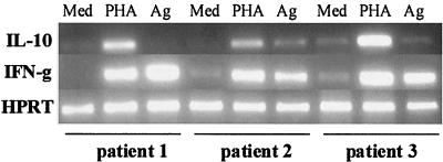
The cytokine profile produced by peripheral blood mononuclear cells (PBMC) in response to leishmania antigens and the ability of interleukin-10 (IL-10) and transforming growth factor beta (TGF-beta) to modulate the immune response were evaluated in 21 mucosal leishmaniasis patients. Patients with mucosal disease exhibited increased gamma interferon (IFN-gamma) and tumor necrosis factor alpha (TNF-alpha) secretion and decreased IL-10 secretion compared to patients with classical cutaneous leishmaniasis. CD4(+) Th1 cells were the main source of IFN-gamma and TNF-alpha production in mucosal leishmaniasis patients. Evaluation of cytokine gene expression in PBMC of these patients showed that there was strong up-regulation of IFN-gamma transcripts upon stimulation with leishmania antigen, in contrast to the baseline levels of IL-10 mRNA. IL-10 suppressed IFN-gamma production by 48% in cell cultures from cutaneous leishmaniasis patients and by 86% in cell cultures from healthy subjects stimulated with purified protein derivative, whereas in similar conditions IL-10 suppressed IFN-gamma production by 19% in cell cultures from mucosal leishmaniasis patients stimulated with leishmania antigen. TGF-beta suppressed IFN-gamma levels to a greater extent in healthy subjects than in mucosal leishmaniasis and cutaneous leishmaniasis patients. These data indicate that a poorly modulated T-cell response in mucosal leishmaniasis patients leads to production of high levels of proinflammatory cytokines, such as IFN-gamma and TNF-alpha, as well as a decreased ability of IL-10 and TGF-beta to modulate this response. These abnormalities may be the basis for the pathological findings observed in this disease.


Figures

FIG. 1.
FIG. 1.
Cytokine profiles in ML and CL patients. IFN-γ and TNF-α production (A) and IL-10 and IL-5 production (B) in PBMC supernatants stimulated with leishmania antigen were determined as described in Materials and Methods. The levels of cytokines were measured by ELISA. Symbols: ▪, ML; ▴, CL.
FIG. 2.
FIG. 2.
Cytokine mRNA expression in PBMC from three ML patients with different stimuli. A total of 3 × 10⁶ PBMC were incubated for 72 h in the presence of medium (Med), PHA, or leishmania antigen (Ag), and then total mRNA was extracted and RT-PCR for IL-10 and IFN-γ were performed to assess cytokine gene expression. Hypoxanthine phosphoribosyltransferase (HPRT) expression was used to normalize samples and for comparison.
FIG. 3.
FIG. 3.
Identification of IFN-γ-producing cells in ML patients. As described in Materials and Methods, CD4+ T cells, CD8+ T cells, or NK cells were removed from PBMC by using magnetic beads. Cultures were stimulated with leishmania antigen for 72 h and assayed for the presence of IFN-γ by ELISA. The data are the means and standard deviations for six patients. The percentages of contamination in the populations with cells removed were as follows: population with CD4+ cells removed, 0.2% CD4+ cells; population with CD8+ cells removed, 2% CD8+ cells; and population with NK cells removed, 1% CD56+ cells. An asterisk indicates that the P value is <0.005.
FIG. 4.
FIG. 4.
Identification of TNF-α-producing cells in ML patients. As described in Materials and Methods, CD4+ T cells or macrophages were removed from PBMC, and the PBMC were stimulated with leishmania antigen. TNF-α-producing cells were identified as described in the legend to Fig. 3. The data are the means and standard deviations for three patients. The percentages of contamination in the populations with cells removed were as follows: population with CD4+ cells removed, 0.2% CD4+ cells; and population with CD8+ cells removed, 2% CD8+ cells. The percentages of esterase-positive cells were 28% ± 3% before removal and 2% ± 0.7% after removal.
FIG. 5.
FIG. 5.
Cell population responsible for TNF-α production following antigenic stimulation. The cellular sources of TNF-α production are shown for two ML patients, as determined by using single-cell cytoplasmic staining and analysis with a flow cytometer. (A) TNF-α production without stimulus. (B and C) Frequency of CD14+ monocytes which produce TNF-α in two independent individuals, one exhibiting a low frequency of TNF-α-producing monocytes (B) and the other exhibiting a higher frequency of TNF-α-producing monocytes (C) following 20 h of culture with SLA. (D and E) TNF-α-producing CD4+ and CD8+ T cells, respectively, from another ML patient following short-term in vitro stimulation with SLA as described in Materials and Methods. TNF-A-PE, TNF-α-phosphatidylethanolamine; CD4-CYCHROM, CD4-Cychrome.
FIG. 6.
FIG. 6.
Regulation of IFN-γ production by IL-10 in ML and CL patients. As described in Materials and Methods, PBMC from ML or CL patients were stimulated with leishmania antigen (La Ag) in the presence or absence of IL-10. IL-10 was used at a concentration of 5 ng/ml. The data are means and standard deviations for seven ML patients and seven CL patients. The percentage of suppression is indicated next to the single asterisk. Two asterisks indicate that the P value is <0.005.

References

    1. Azulay, R. D., and D. R. Azulay, Jr. 1995. Immune-clinical-pathologic spectrum of leishmaniasis. Int. J. Dermatol. 34:303-307. - PubMed
    1. Bacellar, O., A. D'Oliveira, Jr., S. Jeronimo, and E. M. Carvalho. 2000. IL-10 and IL-12 are the main regulatory cytokines in visceral leishmaniasis. Cytokine 12:1228-1231. - PubMed
    1. Barral, A., M. Barral-Netto, E. C. Yong, C. E. Brownell, D. R. Twardzik, and S. G. Reed. 1993. Transforming growth factor beta as a virulence mechanism for Leishmania braziliensis. Proc. Natl. Acad. Sci. USA 90:3442-3446. - PMC - PubMed
    1. Barral-Netto, M., A. Barral, C. Brodskyn, E. M. Carvalho, and S. G. Reed. 1995. Cytotoxicity in human mucosal and cutaneous leishmaniasis. Parasite Immunol. 17:21-28. - PubMed
    1. Barral-Netto, M., A. Barral, C. E. Brownell, Y. A. Skeiky, L. R. Ellingsworth, D. R. Twardzik, and S. G. Reed. 1992. Transforming growth factor-beta in leishmanial infection: a parasite escape mechanism. Science 257:545-548. - PubMed

Publication types

MeSH terms

LinkOut - more resources