. 2002 Dec;70(12):6911-8.
doi: 10.1128/IAI.70.12.6911-6918.2002.

B-cell-deficient mice show an exacerbated inflammatory response in a model of Chlamydophila abortus infection

Affiliations

B-cell-deficient mice show an exacerbated inflammatory response in a model of Chlamydophila abortus infection

Antonio J Buendía et al. Infect Immun. 2002 Dec.

Abstract

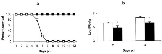
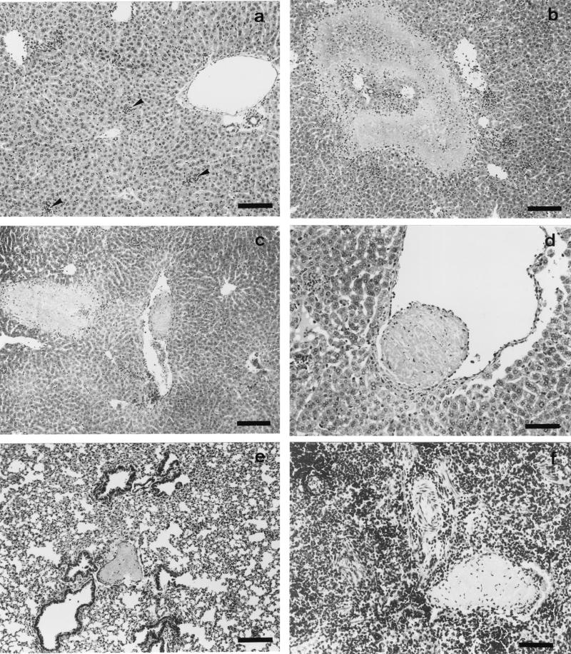
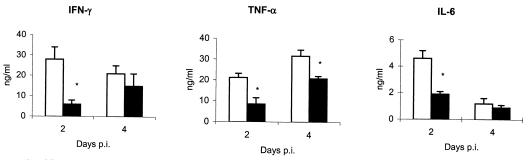
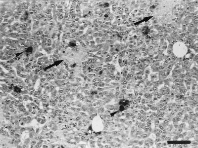
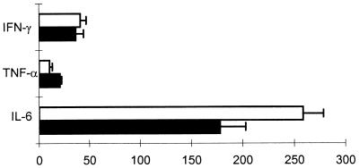
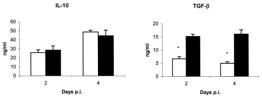
The resolution of Chlamydophila abortus (Chlamydia psittaci serotype 1) infection is dependent on gamma interferon and CD8(+) T cells, and classically, B cells have been considered to play a minimal role in host defense. The role of B cells in the immune response was studied by using a model of infection in mice with genetically modified immunoglobulin M transmembrane domains ( micro MT). In the absence of B cells, infection with C. abortus leads to an acute severe fatal disease that involves a disseminated intravascular coagulation syndrome. micro MT mice displayed an increased level of proinflammatory cytokines in serum, and an increased number of neutrophils was observed in the lesions. The possible deleterious role of neutrophils in the pathogenesis of disease in micro MT mice was determined by depletion of the neutrophils with the monoclonal antibody RB6-8C5. This led to an enhancement of the bacterial burden and early mortality in both micro MT and wild-type mice, while necrotic lesions remained. Analysis of the presence of immunoregulatory cytokines showed significantly lower levels of transforming growth factor beta in the sera of micro MT mice. However, mice lacking mature B cells were able to establish a specific immune response that protected them from a secondary challenge. Taken together, these data suggest an immunomodulatory role for B cells in the early events of C. abortus primary infection that can protect mice against an exaggerated inflammatory response.


Figures

FIG. 1.
FIG. 1.
Evolution of C. abortus infection in μMT and WT mice. (a) Survival of μMT (□) and WT (▪) mice after infection. Groups of eight mice were infected with 106 IFUs of C. abortus. Mice were monitored daily, and mortality rates were calculated. The experiment was repeated twice, and the data shown are the results for two groups of mice of each strain (16 mice per strain). (b) Quantitative isolation of C. abortus on homogenized tissues from livers of μMT (white columns) and WT (black columns) mice collected at days 2 and 4 p.i. The number of inclusions was visualized by indirect immunofluorescence. The results are the means and SD for five mice per strain infected with 106 IFUs of C. abortus. Experiments were performed two more times with similar results. Asterisks indicate significant differences (P < 0.01) between μMT and WT mice at the same day p.i.
FIG. 2.
FIG. 2.
Histopathology of C. abortus-infected μMT and WT mice at day 2 p.i. Paraffin wax sections stained with hematoxylin and eosin show the disseminated intravascular coagulation in μMT mice. (a) Scarce and small inflammatory foci (arrowheads) in the livers of WT mice. (b) Area of coagulative necrosis surrounded by a substantial infiltration of neutrophils in the livers of μMT mice. (c) Thrombotic lesions near an area of coagulative necrosis in the livers of μMT mice. (d) Detail of a thrombotic lesion in the liver. (e and f) Thrombotic lesions in the lungs and spleens, respectively, of μMT mice. Original magnifications, ×100 (a, b, c, and e) and ×200 (d and f); bars, 266 μm (a, b, c, and e) and 133 μm (d and f).
FIG. 3.
FIG. 3.
Levels of the liver-associated enzyme ALT in serum. ALT levels were measured in serum taken from neutrophil-depleted and nondepleted mice at 4 days p.i. The results are expressed as means and SD from five mice per group and are representative of two separate experiments. Asterisks indicate significant differences (P < 0.01) between μMT and WT mice. The double asterisks indicate a significant difference (P < 0.01) between neutrophil-depleted and nondepleted mice of the same strain. Levels of ALT in control serum from uninfected mice were below 10 U/ml.
FIG. 4.
FIG. 4.
Presence of proinflammatory cytokines in the sera of μMT (white columns) and WT (black columns) mice at 2 and 4 days p.i. The results are the means and SD for five mice. Experiments were performed two more times with similar results. Asterisks indicate significant differences (P < 0.01) between μMT and WT mice at the same day p.i.
FIG. 5.
FIG. 5.
Evolution of C. abortus infection in μMT and WT mice depleted of neutrophils by treatment with the MAb RB6-8C5. Nondepleted μMT and WT mice served as controls for the infection study. (a) Survival of neutrophil-depleted (triangles) and nondepleted (squares) μMT (white) and WT (black) mice after infection. Groups of six mice were infected with 106 IFUs of C. abortus. Mice were monitored daily, and mortality rates were calculated. The experiment was repeated twice more, and the data shown are the results for two groups of mice of each strain (12 mice per group). (b) Quantitative isolation of C. abortus on homogenized tissues from livers collected at day 4 p.i. The number of inclusions was visualized by indirect immunofluorescence. The results are the means and SD for five mice. Experiments were performed twice with similar results. The asterisk indicates significant differences (P < 0.01) between neutrophil-depleted and nondepleted mice of the same strain.
FIG. 6.
FIG. 6.
Histopathology and immunoreaction in the livers of neutrophil-depleted μMT mice at day 4 after being infected with C. abortus. The paraffin wax section immunostained by the ABC method shows the distribution of positive immunoreaction in the chlamydial inclusions within hepatocytes (arrowheads) and areas of necrosis (arrows). Notice the lack of inflammatory foci. Original magnification, ×200; bar, 133 μm.
FIG. 7.
FIG. 7.
Administration of antigranulocyte RB6-8C5 MAb during C. abortus infection alters the levels of cytokines in serum. Results shown are the mean percentages and SD of the levels of proinflammatory cytokines in the serum of neutrophil-depleted and nondepleted μMT (white columns) and WT (black columns) mice, with five mice per group. Experiments were performed twice with similar results.
FIG. 8.
FIG. 8.
Presence of immunoregulatory cytokines in the sera of μMT (white columns) and WT (black columns) mice at 2 and 4 days p.i. The results are the means and SD for five mice. Experiments were performed twice with similar results. Asterisks indicate significant differences (P < 0.01) between μMT and WT mice at the same day p.i.

Similar articles

Cited by

References

    1. Bosio, C. M., D. Gradner, and K. L. Elkins. 2000. Infection of B cell-deficient mice with CDC 1551, a clinical isolate of Mycobacterium tuberculosis: delay in dissemination and development of lung pathology. J. Immunol. 164:6417-6425. - PubMed
    1. Bosio, C. M., and K. L. Elkins. 2001. Susceptibility to secondary Francisella tularensis live vaccine strain infection in B-cell-deficient mice is associated with neutrophilia but not with defects in specific T-cell-mediated immunity. Infect. Immun. 69:194-203. - PMC - PubMed
    1. Buendía, A. J., R. Montes de Oca, J. A. Navarro, J. Sánchez, F. Cuello, and J. Salinas. 1999. Role of polymorphonuclear neutrophils in a murine model of Chlamydia psittaci-induced abortion. Infect. Immun. 67:2110-2116. - PMC - PubMed
    1. Buendía, A. J., J. Salinas, J. Sánchez, M. C. Gallego, A. Rodolakis, and F. Cuello. 1997. Localisation by immunoelectron microscopy of antigens of Chlamydia psittaci suitable for diagnosis or vaccine development. FEMS Microbiol. Lett. 150:113-119. - PubMed
    1. Buendía, A. J., J. Sánchez, L. Del Río, B. Garcés, M. C. Gallego, M. R. Caro, A. Bernabé, and J. Salinas. 1999. Differences in the immune response against ruminant chlamydial strains in a murine model. Vet. Res. 30:495-507. - PubMed

Publication types