. 2002 Dec;70(12):6976-86.
doi: 10.1128/IAI.70.12.6976-6986.2002.

Expression of a novel Leishmania gene encoding a histone H1-like protein in Leishmania major modulates parasite infectivity in vitro

Affiliations

Expression of a novel Leishmania gene encoding a histone H1-like protein in Leishmania major modulates parasite infectivity in vitro

Fotini T Papageorgiou et al. Infect Immun. 2002 Dec.

Abstract

We describe identification and characterization of a novel two-copy gene of the parasitic protozoan Leishmania that encodes a nuclear protein designated LNP18. This protein is highly conserved in the genus Leishmania, and it is developmentally regulated. It is an alanine- and lysine-rich protein with potential bipartite nuclear targeting sequence sites. LNP18 shows sequence similarity to H1 histones of trypanosomatids and of higher eukaryotes and in particular with histone H1 of Leishmania major. The nuclear localization of LNP18 was determined by indirect immunofluorescence and Western blot analysis of isolated nuclei by using antibodies raised against the recombinant protein as probes. The antibodies recognized predominantly a 18-kDa band or a 18-kDa-16-kDa doublet. Photochemical cross-linking of intact parasites followed by Western blot analysis provided evidence that LNP18 is indeed a DNA-binding protein. Generation of transfectants overexpressing LNP18 allowed us to determine the role of this protein in Leishmania infection of macrophages in vitro. These studies revealed that transfectants overexpressing LNP18 are significantly less infective than transfectants with the vector alone and suggested that the level of LNP18 expression modulates Leishmania infectivity, as assessed in vitro.


Figures

FIG. 1.
FIG. 1.
Nucleotide sequence of L. major LNP18 cDNA. The deduced amino acid sequence is indicated below the DNA sequence. The domain that contains the eight potential sites of bipartite nuclear sequences (bipartite nuclear localization signals) is underlined. The amino acid sequence is indicated by the single-letter amino acid code.
FIG. 2.
FIG. 2.
(A) Alignment of amino acid sequences of trypanosomatid histones H1 and LNP18. (B) Alignment of LNP18 and L. major histone H1 (19) sequences. The asterisks, colons, and dots indicate aligned residues that are identical, very similar, and similar, respectively. Gaps (dashes) were introduced for optimal alignment. The numbers at the beginning and the end of a line indicate amino acid positions in the parent proteins. L.brazil., L. braziliensis; L.mexic., L. mexicana.
FIG. 3.
FIG. 3.
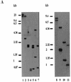
(A) Southern blots of L. major (lanes 1 to 7) and L. infantum (lanes 8 to 11) genomic DNAs digested with various restriction enzymes. Blots were hybridized with LNP18 cDNA. The following restriction enzymes were used: HindIII (lanes 1 and 8); HindIII and EcoRI (lane 2); BglI (lanes 3 and 9); BglI and AvaI (lane 4); AvaI (lane 5); Eco0109I (lanes 6 and 10); and BglI and Eco0109I (lanes 7 and 11). (B) Map I is a physical map of L. major H1 (copies sw3.0 and sw3.1) and the L. infantum LNP18 gene (EMBL/GenBank/DDBJ accession no. AJ223860, AJ223861, and AF469106, respectively). Map II is a physical map of the sequenced gene of L. infantum and of the two copies of the L. infantum LNP18 gene deduced from both Southern blot analysis (panel A, lanes 8 to 11) and restriction enzyme analysis of the sequenced LNP18 DNA gene. Map III is a physical map of the two copies of L. major LNP18 deduced from Southern blot analysis (panel A, lanes 1 to 7) and restriction enzyme analysis of the sequenced LNP18 cDNA. The cross-hatched boxes indicate the homology of sw3.1 and sw3.0 to the cDNA of L. major LNP18. The solid boxes correspond to the LNP18 cDNA sequence in both species. The dashed lines represent DNA that has not been sequenced yet. Enzyme abbreviations: E, EarI; A, AvaI; B, BglI; Ec, Eco0109I.
FIG. 3.
FIG. 3.
(A) Southern blots of L. major (lanes 1 to 7) and L. infantum (lanes 8 to 11) genomic DNAs digested with various restriction enzymes. Blots were hybridized with LNP18 cDNA. The following restriction enzymes were used: HindIII (lanes 1 and 8); HindIII and EcoRI (lane 2); BglI (lanes 3 and 9); BglI and AvaI (lane 4); AvaI (lane 5); Eco0109I (lanes 6 and 10); and BglI and Eco0109I (lanes 7 and 11). (B) Map I is a physical map of L. major H1 (copies sw3.0 and sw3.1) and the L. infantum LNP18 gene (EMBL/GenBank/DDBJ accession no. AJ223860, AJ223861, and AF469106, respectively). Map II is a physical map of the sequenced gene of L. infantum and of the two copies of the L. infantum LNP18 gene deduced from both Southern blot analysis (panel A, lanes 8 to 11) and restriction enzyme analysis of the sequenced LNP18 DNA gene. Map III is a physical map of the two copies of L. major LNP18 deduced from Southern blot analysis (panel A, lanes 1 to 7) and restriction enzyme analysis of the sequenced LNP18 cDNA. The cross-hatched boxes indicate the homology of sw3.1 and sw3.0 to the cDNA of L. major LNP18. The solid boxes correspond to the LNP18 cDNA sequence in both species. The dashed lines represent DNA that has not been sequenced yet. Enzyme abbreviations: E, EarI; A, AvaI; B, BglI; Ec, Eco0109I.
FIG. 4.
FIG. 4.
(A) Northern blot analysis of total RNAs extracted from promastigotes of different species of Leishmania (L. amazonensis, L. tropica, L. donovani, and L. major [lanes1 to 3 and 5, respectively]) and L. major amastigotes (lane 4), separated on a 1.2% (wt/vol) agarose-formaldehyde gel. After blotting, the filters were hybridized either with LNP18 cDNA and the β-tubulin probe (lanes 1 to 3) or with LNP18 cDNA and the gp63 probe (lanes 4 and 5) and autoradiographed. The marker on the right indicates the size and mobility of 28Sβ rRNA. (B) RT-PCR analysis of amastigotes and promastigotes. Total RNA isolated from promastigotes (lanes 1 and 2) or amastigotes (lanes 3 and 4) was reversed transcribed, and primers derived from LNP18 (lanes 2 and 4) and β-actin (lanes 1 and 3) were used. The positions of size markers are indicated on the left.
FIG. 5.
FIG. 5.
(A) Gel electrophoresis and immunoblotting of L. major, L. infantum, and L. amazonensis promastigotes (lanes 1 to 3, respectively) probed with antibodies raised against the recombinant protein (anti-rLNP18). The positions of molecular size standards are indicated on the left. (B) Western blot analysis of lysates from lesion-derived amastigotes (lane 1) and promastigotes (lane 2). Lysates were separated by SDS-15% PAGE, electroblotted, and probed with anti-rLNP18 and α-tubulin antibodies (NeoMarkers). The positions of molecular size standards are indicated on the left. (C) Western blot analysis of L. major promastigotes grown to the early logarithmic phase (day 3) (lane 2) and the stationary phase (day 5) (lane 1). The blot was probed with anti-rLNP18 and α-tubulin antibodies. (D) L. major isolated nuclei and cytoplasmic extracts from the same number of parasites (lanes 1 and 2, respectively). SDS-PAGE was performed under reducing conditions on 12.5% polyacrylamide gels. The positions of molecular size standards are indicated on the left. (E and F) Immunofluorescence labeling of promastigotes when the anti-rLNP18 antibodies were used as probes. L. major promastigotes (1.5 × 10 6 cells/ml of PBS) were fixed on glass microscope slides. Binding of anti-rLNP18 in the nucleus was visualized by using a secondary antibody conjugated with fluorescein isothiocyanate (FITC) and either phase-contrast microscopy performed with a photomicroscope (E) or confocal laser microscopy (F). For confocal microscopy an overlay image is also shown.
FIG. 5.
FIG. 5.
(A) Gel electrophoresis and immunoblotting of L. major, L. infantum, and L. amazonensis promastigotes (lanes 1 to 3, respectively) probed with antibodies raised against the recombinant protein (anti-rLNP18). The positions of molecular size standards are indicated on the left. (B) Western blot analysis of lysates from lesion-derived amastigotes (lane 1) and promastigotes (lane 2). Lysates were separated by SDS-15% PAGE, electroblotted, and probed with anti-rLNP18 and α-tubulin antibodies (NeoMarkers). The positions of molecular size standards are indicated on the left. (C) Western blot analysis of L. major promastigotes grown to the early logarithmic phase (day 3) (lane 2) and the stationary phase (day 5) (lane 1). The blot was probed with anti-rLNP18 and α-tubulin antibodies. (D) L. major isolated nuclei and cytoplasmic extracts from the same number of parasites (lanes 1 and 2, respectively). SDS-PAGE was performed under reducing conditions on 12.5% polyacrylamide gels. The positions of molecular size standards are indicated on the left. (E and F) Immunofluorescence labeling of promastigotes when the anti-rLNP18 antibodies were used as probes. L. major promastigotes (1.5 × 10 6 cells/ml of PBS) were fixed on glass microscope slides. Binding of anti-rLNP18 in the nucleus was visualized by using a secondary antibody conjugated with fluorescein isothiocyanate (FITC) and either phase-contrast microscopy performed with a photomicroscope (E) or confocal laser microscopy (F). For confocal microscopy an overlay image is also shown.
FIG. 6.
FIG. 6.
Monitoring LNP18 expression in transfected LV39 parasites. (A) Southern blot analysis of extrachromosomal DNA isolated from parasites transfected with pX63-HYG and from parasites transfected with pX63-HYG-LNP18 (lanes 1 and 3 and lanes 2 and 4, respectively). The DNA (5 μg per lane) was digested with BglI and probed with the BamHI-EcoRI fragment of the HYG coding region (lanes 1 and 2). The membrane was stripped and reprobed with the LNP18 coding region (lanes 3 and 4). The positions of size markers are indicated on the left. (B) RT-PCR analysis of the transfectants. Total RNA extracted from parasites transfected with either pX63-HYG-LNP18 (lanes 1and 2) or pX63-HYG (lanes 3 and 4) was reversed transcribed by using the LNP18-derived primers (lanes 2 and 4) and the β-actin-derived primers (lanes 1 and 3), which were used as a control. The positions of size markers are indicated on the left. (C) Immunoblot analysis of transfected LV39 total lysates (lane1, pX63-HYG; lane 2, pX63-HYG-LNP18). The blot was probed with anti-LNP18 and anti-α-tubulin antibodies (used as a loading control). The positions of standard size markers are indicated on the left.
FIG. 7.
FIG. 7.
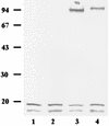
In vitro UV-cross-linking analysis of nuclear L. major extracts. UV irradiation and Western blot analysis of the extracts were performed as described in Materials and Methods. Lane 1, DNase I treatment prior to UV treatment; lane 2, nontreated extracts used as a control; lane 3, UV treatment followed by DNase I treatment; lane 4, UV treatment.
FIG. 8.
FIG. 8.
In vitro infection assays. (A) Macrophages were infected with wild-type lesion-derived promastigotes and LV39 transfected with pX63-HYG and with pX63-HYG-LNP18 for 5 h, washed, and incubated for 72 h (lanes 1 to 3, respectively). The cultures were then washed with medium and lysed with 0.01% (wt/vol) SDS to release the surviving amastigotes, which were cultured for an additional 48 h. Cultures were subsequently pulsed with 1 μCi of [3H]thymidine per well, and incorporation of radioactivity was counted with a β-counter. Data from three independent experiments (12 wells per transfectant or wild-type promastigotes) were pooled, and the standard errors are indicated by error bars. (B) Fluorescence-activated cell sorter histograms acquired with noninfected macrophages (histogram 1) and macrophages infected with LV39 wild type (histogram 2) and LV39 transfected with either pX63-HYG (histogram 3) or pX63-HYG-LNP18 (histogram 4). The macrophages were infected for 5 h, washed, and incubated for 72 h. The cultures were washed again and processed for fluorescence-activated cell sorter analysis as described in Materials and Methods.

Similar articles

Cited by

References

    1. Arents, G., R. W. Burlingame, B.-C. Wang, M. E. Love, and E. Moudrianakis. 1991. The nucleosomal core histone octamer at 3.1 Å resolution: a tripartite protein assembly and a left-handed superhelix. Proc. Natl. Acad. Sci. USA 88:10148-10152. - PMC - PubMed
    1. Arents, G., and E. N. Moudrianakis. 1995. The histone fold: a ubiquitous architectural motif utilized in DNA compaction and protein dimerization. Proc. Natl. Acad. Sci. USA 92:11170-11174. - PMC - PubMed
    1. Aslund, L., L. Carisson, J. Henriksson, M. Rydaker, C. Toro, N. Galanti, and U. Pettersson. 1994. A gene family encoding heterogeneous histone H1 proteins in Trypanosoma cruzi. Mol. Biochem. Parasitol. 65:317-330. - PubMed
    1. Astolfi, F. S., C. Martins de Sa, and E. S. Gander. 1980. On the chromatin structure of Trypanosoma cruzi. Mol. Biochem. Parasitol. 1:45-53. - PubMed
    1. Bader, T., and J. Wietzerbin. 1994. Nuclear accumulation of interferon gamma. Proc. Natl. Acad. Sci. USA 91:11831-11835. - PMC - PubMed

Publication types

MeSH terms

LinkOut - more resources