. 2002 Nov;110(10):1515-23.
doi: 10.1172/JCI15564.

IFN-gamma protects short-term ovarian carcinoma cell lines from CTL lysis via a CD94/NKG2A-dependent mechanism

Affiliations

IFN-gamma protects short-term ovarian carcinoma cell lines from CTL lysis via a CD94/NKG2A-dependent mechanism

Karl-Johan Malmberg et al. J Clin Invest. 2002 Nov.

Abstract

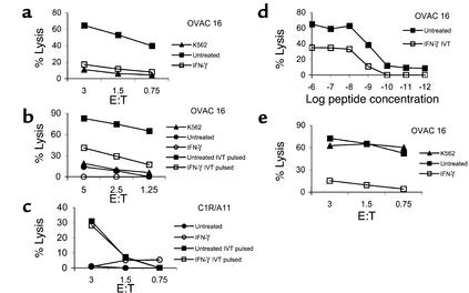
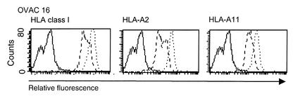
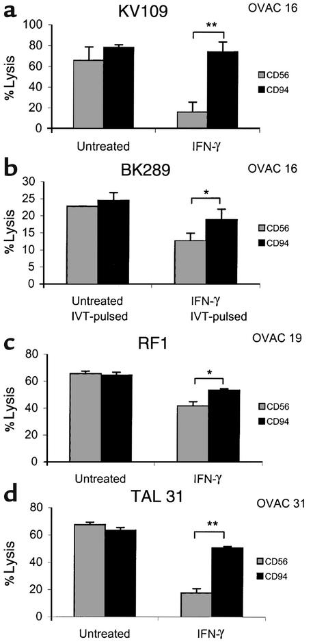
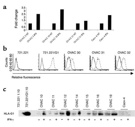
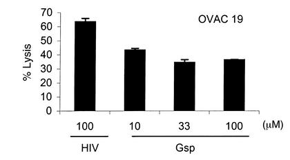
IFN-gamma regulates the immunogenicity of target cells by increasing their expression of HLA class I molecules. This facilitates the T cell receptor-mediated recognition by CD8(+) T cells but decreases target cell sensitivity to lysis by NK cells due to engagement of inhibitory NK receptors. In this study, short-term tumor cell lines from patients with advanced ovarian carcinomas were established. We demonstrate the paradoxical finding that IFN-gamma treatment of these short-term ovarian carcinoma cell lines (OVACs) resulted in resistance of tumor cells to lysis by peptide- and allospecific CD8(+) T cells. Blocking experiments revealed that this phenomenon was dependent on enhanced inhibitory signalling via CD94/NKG2A receptors expressed on the effector cells. This was associated with increased expression of HLA-E mRNA and HLA-G at the protein level in IFN-gamma-treated OVACs. Furthermore, pulsing of untreated OVACs with the leader sequence peptide of HLA-G protected these cells from lysis by CTLs, thus mimicking the inhibitory effect of IFN-gamma. This study provides evidence that CD94/NKG2A receptors play an important role in regulating T cell activity against tumors and shows that IFN-gamma modulation of target cells may shift the balance of triggering and inhibitory signals to T cells, turning off their cytolytic activity.


Figures

Figure 1
Figure 1
IFN-γ protects short-term ovarian carcinomas from CTL lysis. The effect of IFN-γ on CTL recognition of OVACs was evaluated in 4-hour 51Cr-release assays. (a) K562 and untreated or IFN-γ–treated OVAC 16 (HLA-A11+) were used as targets for the HLA-A11–specific, CD8+ CTL clone KV109. (b) OVAC 16 was untreated or treated with IFN-γ and then pulsed or not pulsed with the HLA-A11–restricted, EBNA4-derived epitope IVT at a concentration of 1 μg/ml and used as a target for the IVT-specific, CD8+ T cell clone BK289. (c) C1R/A11 was untreated or treated with IFN-γ and then pulsed or not pulsed with the IVT peptide (1 μg/ml) and used as a target for BK289. (d) Untreated or IFN-γ–treated OVAC 16 was prepulsed with IVT peptide at concentrations ranging from 10–12 to 10–6 g/ml and used as a target for BK289 at an effector-to-target ratio of 3:1. Recognition of non-pulsed targets was below 7%. Results from one of at least three experiments are shown. (e) K562 and untreated or IFN-γ–treated OVAC 16 was used as a target for autologous TALs.
Figure 2
Figure 2
Intact regulatory function of IFN-γ on HLA class I expression in OVACs. OVAC 16 was untreated or treated with IFN-γ for 48 hours and then stained for the expression of total HLA class I, HLA-A2, and HLA-A11. Stainings of untreated cells (dashed line) and IFN-γ–treated cells (dotted line) are shown. Isotype control stainings are depicted as a solid line. Histograms from one representative staining out of three of this particular tumor is shown.
Figure 3
Figure 3
Blockade of CD94/NKG2A inhibitory receptors restores killing of IFN-γ–treated OVACs. The effect of anti-CD56 (MOC-1) or anti-CD94 (HP3D9) mAb’s on the IFN-γ–mediated protection of OVACs was evaluated in the following situations: The HLA-A11+ OVAC 16 was used as target for the HLA-A11–specific T cell clone KV109 (a) and for the IVT-specific T cell clone BK289 (b). The peptide concentration in b was 1 μg/ml. Recognition of unpulsed targets by BK289 was below 5% and is not shown. (c) The HLA class I–restricted, tumor-reactive CTL line RF1 was used as an effector against untreated and IFN-γ–treated OVAC 19. (d) Reactivity of TAL 31 against autologous OVAC 31. Error bars represent SD of triplicate cultures. *P < 0.05, **P < 0.01 as determined by two-tailed Student t test.
Figure 4
Figure 4
Expression of nonclassical HLA molecules. (a) The expression of HLA-E mRNA was evaluated in indicated OVACs by real-time quantitative PCR assay. Results are presented as fold increase in IFN-γ–treated cells (IFN) compared with untreated cells (NT). Results from one of two experiments are shown. (b) The expression of HLA-G by freshly isolated ovarian tumor cells was monitored by flow cytometry analysis. Indicated cells were stained with isotype control (mouse IgG2a) (solid line) or anti–HLA-G1 (87G) (dotted line). Results from one representative staining of at least ten are shown. (c) Western blot analysis of HLA-G expression in indicated OVACs and Caov-4 was performed using the HLA-G–specific mAb MEM-G/1 at a concentration of 1 μg/ml. The LCLs 721.221 and 721.221 transfected with HLA-G1 (721.221/G1) were used as positive and negative controls. These were diluted 1:10 because of the excess amount of HLA-G expressed by the 721.221/G1 transfectant. Cell extracts of untreated and IFN-γ–treated samples were loaded in equal amounts as determined by Western blots for β-actin (data not shown).
Figure 5
Figure 5
Role of the HLA-G leader sequence peptide. OVAC 19 was incubated with different concentrations of Gsp as indicated in the figure. HLA-A2–binding HIV peptide (HIV-RT 476-484) was used as negative control at 100 μg/ml. HIV- and Gsp-pulsed OVAC 19 was then used as target in a 4-hour 51Cr-release assay. The HLA class I–restricted, tumor-specific CTL line RF1 was used as effector. Results from one of two experiments are shown.

Comment in

References

    1. Shankaran V, et al. IFNgamma and lymphocytes prevent primary tumour development and shape tumour immunogenicity. Nature. 2001;410:1107–1111. - PubMed
    1. Kaplan DH, et al. Demonstration of an interferon gamma-dependent tumor surveillance system in immunocompetent mice. Proc Natl Acad Sci USA. 1998;95:7556–7561. - PMC - PubMed
    1. Dighe AS, Richards E, Old LJ, Schreiber RD. Enhanced in vivo growth and resistance to rejection of tumor cells expressing dominant negative IFN gamma receptors. Immunity. 1994;1:447–456. - PubMed
    1. Rock KL, Goldberg AL. Degradation of cell proteins and the generation of MHC class I-presented peptides. Annu Rev Immunol. 1999;17:739–779. - PubMed
    1. Moretta A, et al. Receptors for HLA class-I molecules in human natural killer cells. Annu Rev Immunol. 1996;14:619–648. - PubMed

Publication types

MeSH terms

LinkOut - more resources