. 2002 Nov;110(10):1549-57.
doi: 10.1172/JCI15454.

Modulation of tumor growth by inhibitory Fc(gamma) receptor expressed by human melanoma cells

Affiliations

Modulation of tumor growth by inhibitory Fc(gamma) receptor expressed by human melanoma cells

Lydie Cassard et al. J Clin Invest. 2002 Nov.

Abstract

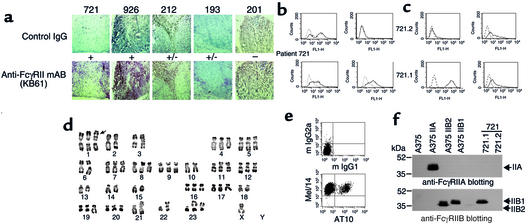
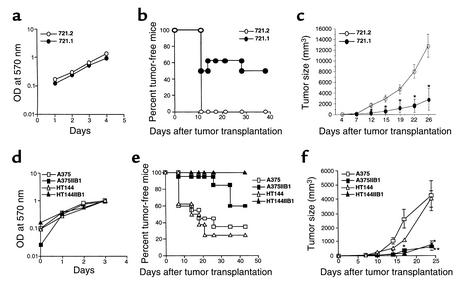
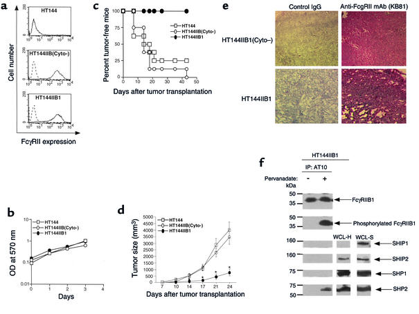
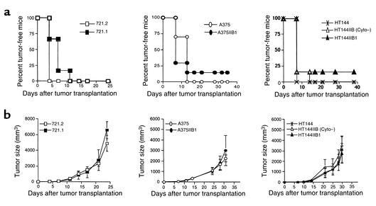
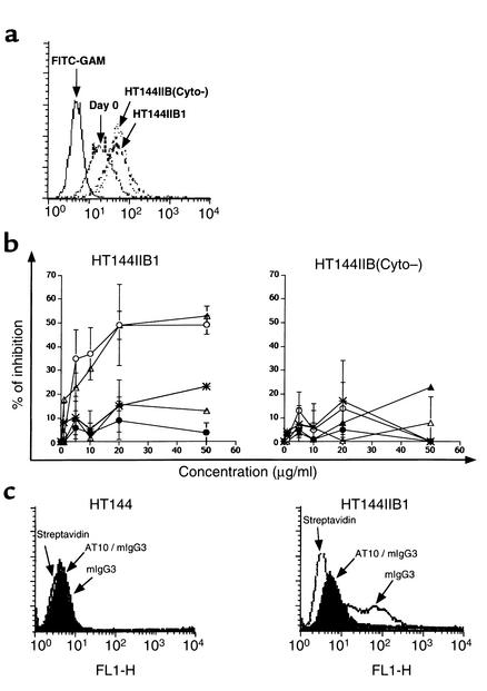
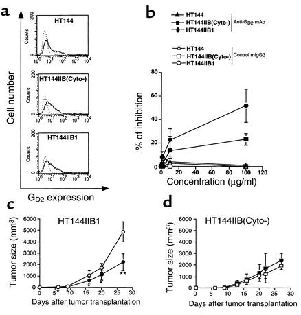
The efficacy of anti-tumor IgG reflects the balance between opposing signals mediated by activating and inhibitory Fc(gamma) receptors (Fc(gamma)Rs) expressed by effector cells. Here, we show that human malignant melanoma cells express the inhibitory low-affinity Fc(gamma) receptor Fc(gamma)RIIB1 in 40% of tested metastases. When melanoma cells were grafted in nude mice, a profound inhibition of Fc(gamma)RIIB1 tumor growth that required the intracytoplasmic region of the receptor was observed. IgG immune complexes (ICs) may be required for this inhibition, since sera from nude mice bearing tumors contained IgG that decreased the proliferation of Fc(gamma)RIIB1-positive cells in vitro, and tumor development of Fc(gamma)RIIB1-positive melanoma lines was not inhibited in antibody-defective severe combined immunodeficiency (SCID) mice. Passive immunization of SCID mice with anti-ganglioside G(D2) antibody resulted in significant inhibition of growth of Fc(gamma)RIIB1-positive tumors in an intracytoplasmic-dependent manner. Altogether, these data suggest that human melanoma cells express biologically active inhibitory Fc(gamma)RIIB1, which regulates their development upon direct interaction with anti-tumor antibodies. Therefore, Fc(gamma)R expression on human tumors may be one component of the efficacy of antibody-mediated therapies, and Fc(gamma)R-positive tumors could be the most sensitive candidates for such treatments.


Figures

Figure 1
Figure 1
Human metastatic melanomas express inhibitory FcγRII. (a) Immunohistochemical analysis using anti-FcγRII mAb KB61 in liver (patients 721, 926, and 212), lymph node (patient 193), or subcutaneous (patient 201) metastases from patients with cutaneous (patients 926, 193, and 201) or uveal (patients 212 and 721) melanoma (bottom). No immunostaining was detected when primary antibody was replaced with isotype control (top). ×30. (b and c) Cell line derived from patient 721 expresses FcγRII. Cells from this melanoma line (721 and its derivative clones, 721.1 and 721.2) were incubated with anti-FcγRIIA+B (AT10) or anti-FcγRIIA (IV.3) mAb’s, anti-melanoma mAb’s (GD2 mAb’s), anti–HLA-DR mAb, or control isotype (dotted histograms). (d and e) Cell line derived from patient 721 is a melanoma cell line. (d) R-banded karyotype of 721 cell line. Numerous structural and numerical chromosomal aberrations are present. See the abnormality of 1p (arrow), probably involving 3p. (e) Double staining was performed by incubating cells with AT10 and anti-melanoma marker (Mel/14) mAb’s (bottom) or with control isotypes (top). (f) Analysis of the FcγRII isoforms expressed by the 721 cell line, by immunoprecipitation of FcγRII with AT10 in cell lysates from 721.1, 721.2, A375, A375IIB1, A375IIB2, and A375IIA cell lines, and by immunoblotting with anti-FcγRIIA (260) (top panel) or anti-FcγRIIB/IC (bottom panel) polyclonal antibodies. Molecular weight standards (kDa) are shown in the left lane. mIgG, mouse IgG.
Figure 2
Figure 2
FcγRIIB1 expression is associated with inhibition of development of human melanoma tumors in nude mice. (a and d) In vitro proliferation of FcγRIIB1-negative (721.2, A375, or HT144) and FcγRIIB1-positive (721.1, A375IIB1, or HT144IIB1 transfectants) melanoma lines, determined by an MTT assay on days 1–4. (b and e) Tumor uptake in nude mice, determined by subcutaneous inoculation of 2 × 10⁶ 721.1 and 721.2 cells (b) or 1 × 10⁶ A375, HT144, A375IIB1, or HT144IIB1 cells (e). Results are expressed as percentage of mice (n = 30) that remained tumor-free after challenge in three experiments. (c and f) Tumor growth, measured by volume after injection of 4 × 10⁶ 721.1 and 721.2 cells (c) or 2 × 10⁶ A375, HT144, A375IIB1, or HT144IIB1 cells (f). Ten mice were used per experiment in each group, and mean tumor volume values, with SD, of mice bearing tumor are shown. Data are representative of three independent experiments. *P < 0.03, **P < 0.002 between FcγRIIB1-positive and FcγRIIB1-negative tumors.
Figure 3
Figure 3
The FcγRIIB1-mediated inhibition of melanoma development in nude mice requires the receptor’s cytoplasmic tail. HT144IIB1 and HT144IIB(Cyto) cell lines were used. (a) Expression of FcγRIIB1 as assessed the day before injection by flow cytometry using AT10 or control IgG1 (dotted histograms). (b) In vitro proliferations of melanoma lines were determined by an MTT assay on days 1–4. (c) Tumor uptake incidence as determined by subcutaneous inoculation of nude mice with 1 × 10⁶ cells. Results are expressed as percentage of mice (n = 20) that remained tumor-free after challenge in two experiments. (d) Tumor growth as measured by volume after inoculation of 2 × 10⁶ cells. Ten mice were used per experiment in each group, and mean tumor volume values of mice bearing tumor with SDs are shown. Data are representative of two independent experiments. *P < 0.01 between HT144IIB1 tumors and HT144 or HT144IIB(Cyto) tumors. (e) Immunohistochemical analysis of FcγRII expression using KB61 mAb (right panels), on tumor tissues at day 21 after injection in nude mice of HT144IIB1 (bottom) or HT144IIB(Cyto) (top) cells. ×30. (f) Recruitment of SHP2 phosphatase to phosphorylated FcγRIIB1. HT144IIB1 cells (6 × 10⁷) were treated or not treated with 100 μM pervanadate for 10 minutes. Cells were lysed and FcγRIIB was immunoprecipitated using AT10 mAb. Immunoprecipitated materials were electrophoresed and Western blotted with anti-FcγRIIB/IC, anti-phosphotyrosine, anti-SHIP1, anti-SHIP2, anti-SHP1, and anti-SHP2 Ab’s. Whole cell lysates of HT144IIB1 (WCL-H) or ST486 (WCL-S) were used as positive controls.
Figure 4
Figure 4
The development of human melanomas is independent of FcγRIIB1 expression in SCID mice. (a) Tumor uptake, determined by inoculation of 2 × 10⁶ 721.1 or 721.2 cells or 1 × 10⁶ A375, A375IIB1, HT144, HT144IIB(Cyto), or HT144IIB1 cells in SCID mice. Results are expressed as percentage of mice (n = 18) that remained tumor-free after challenge in two experiments. (b) Tumor growth, measured by volume after inoculation of 4 × 10⁶ 721.1 or 721.2 cells or 2 × 10⁶ A375, A375IIB1, HT144, HT144IIB(Cyto), or HT144IIB1 cells. Nine mice were used per experiment in each group, and mean tumor volume values, with SD, of mice bearing tumor are shown. Data are representative of two independent experiments.
Figure 5
Figure 5
Sera from nude mice contain IgG antibodies that bind to melanoma cells and inhibit proliferation of FcγRIIB1-positive melanoma cells in an intracytoplasmic-dependent manner. (a) Day-0 and day-14 sera from nude mice bearing HT144IIB(Cyto) or HT144IIB1 tumors, or medium alone (FITC-GAM), were incubated with HT144 cells, and binding of IgG was assessed by flow cytometry. (b) HT144IIB1 (left panel)or HT144IIB(Cyto) (right panel) cells (5 × 10³/well) were incubated with the indicated concentrations of IgG isolated from sera of nude mice at day 0 (cross) or bearing day-14 HT144IIB1 (open circles) or HT144IIB(Cyto) (open triangles) tumors or with the same volume of control eluates from sera of SCID mice bearing day-14 HT144IIB1 (filled circles) or HT144IIB(Cyto) (filled triangles) tumors for 24 hours to FCS-free medium. This was followed by a 24-hour pulse with 3H-thymidine. The percentages of inhibition were estimated with the formula [(1 – cpm of stimulated cells / cpm of unstimulated cells) × 100]. Each point represents results from three independent experiments performed in triplicate (mean ± SD). (c) HT144 (left panel) or HT144IIB1 (right panel) cells were preincubated with AT10, then with biotin-labeled mouse IgG3, and then with FITC-conjugated streptavidin, or were not preincubated. Binding of mouse IgG3 was compared with background binding of FITC-conjugated streptavidin.
Figure 6
Figure 6
Anti-GD2 mAb inhibits growth of FcγRIIB1-positive tumor cells in vitro and in vivo, in an intracytoplasmic-dependent manner. (a) Expression of GD2 on cells as assessed by flow cytometry using 7A4 or a control isotype (dotted histograms). (b) Inhibition of proliferation of melanoma cells in vitro. Cells (5 × 10³ cells per well) were incubated with the indicated concentrations of anti-GD2 mAb 7A4 (filled symbols) or control mouse IgG3 (open symbols). 3H-thymidine incorporation rate and percentages of inhibition were measured as described in Figure 5. Each point represents results from three independent experiments performed in triplicate (mean ± SD). (c and d) Inhibition of growth of HT144IIB1 but not of HT144IIB(Cyto) tumors in SCID mice injected intravenously with anti-GD2 mAb. SCID mice were inoculated subcutaneously with 2 × 10⁶ HT144IIB1 (c) and HT144IIB(Cyto) (d) cells, followed by biweekly intravenous injections of 50 μg of mAb 7A4 (filled symbols) or PBS (open symbols). Eight mice were used per experiment in each group, and mean tumor volume values, with SD, of mice bearing tumor are shown. *P < 0.05, **P < 0.0001.

References

    1. Ravetch JV, Bolland S. IgG Fc receptors. Annu Rev Immunol. 2001;19:275–290. - PubMed
    1. Amigorena S, et al. Cytoplasmic domain heterogeneity and functions of IgG Fc receptors in B lymphocytes. Science. 1992;256:1808–1812. - PubMed
    1. Van den Herik-Oudijk IE, Capel PJ, van der Bruggen T, Van de Winkel JG. Identification of signaling motifs within human Fc gamma RIIa and Fc gamma RIIb isoforms. Blood. 1995;85:2202–2211. - PubMed
    1. Fridman WH, et al. Structural bases of Fc gamma receptor functions. Immunol Rev. 1992;125:49–76. - PubMed
    1. White CA, Weaver RL, Grillo-Lopez AJ. Antibody-targeted immunotherapy for treatment of malignancy. Annu Rev Med. 2001;52:125–145. - PubMed

Publication types