Virus-specific CD8+ lymphocytes share the same effector-memory phenotype but exhibit functional differences in acute hepatitis B and C
- PMID: 12438568
- PMCID: PMC136708
- DOI: 10.1128/jvi.76.24.12423-12434.2002
Virus-specific CD8+ lymphocytes share the same effector-memory phenotype but exhibit functional differences in acute hepatitis B and C
Abstract
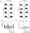
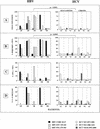
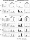
Hepatitis B and hepatitis C viruses (HBV and HCV) are both noncytopathic and can cause acute and chronic infections of the liver. Although they share tropism for the same organ, development of chronic hepatitis is much more frequent following HCV infection, suggesting different mechanisms of viral persistence. In this study, we show that circulating HBV- and HCV-specific tetramer-positive CD8 cells during the acute phase of hepatitis B and C belong almost entirely to an effector-memory subset (CCR7(-) CD45RA(-)). Despite this phenotypic similarity, HBV- and HCV-specific CD8 cells show striking functional differences. HBV-specific tetramer-positive CD8 cells express high perforin content ex vivo, expand vigorously, and display efficient cytotoxic activity and gamma interferon (IFN-gamma) production upon peptide stimulation. A comparable degree of functional efficiency is maintained after the resolution of hepatitis B. In contrast, HCV-specific CD8 cells in the acute phase of hepatitis C express significantly lower levels of perforin molecules ex vivo and show depressed CD8 function in terms of proliferation, lytic activity, and IFN-gamma production, irrespective of the final outcome of the disease. This defect is transient, because HCV-specific CD8 cells can progressively improve their function in patients with self-limited hepatitis C, while the CD8 function remains persistently depressed in subjects with a chronic evolution.
Figures


Similar articles
-
Dynamic decrease in PD-1 expression correlates with HBV-specific memory CD8 T-cell development in acute self-limited hepatitis B patients.J Hepatol. 2009 Jun;50(6):1163-73. doi: 10.1016/j.jhep.2009.01.026. Epub 2009 Mar 29. J Hepatol. 2009. PMID: 19395117
-
Visual demonstration of hepatitis C virus-specific memory CD8(+) T-cell expansion in patients with acute hepatitis C.Hepatology. 2001 Jan;33(1):287-94. doi: 10.1053/jhep.2001.21164. Hepatology. 2001. PMID: 11124847
-
Dysfunction and functional restoration of HCV-specific CD8 responses in chronic hepatitis C virus infection.Hepatology. 2007 Mar;45(3):588-601. doi: 10.1002/hep.21541. Hepatology. 2007. PMID: 17326153
-
CD8+ T-Cell Responses in Hepatitis B and C: The (HLA-) A, B, and C of Hepatitis B and C.Dig Dis. 2016;34(4):396-409. doi: 10.1159/000444555. Epub 2016 May 11. Dig Dis. 2016. PMID: 27170395 Review.
-
[The role of the virus specific T-cell response in acute and chronic HBV and HCV infection].Z Gastroenterol. 2004 Jan;42(1):39-46. doi: 10.1055/s-2004-812688. Z Gastroenterol. 2004. PMID: 14997402 Review. German.
Cited by
-
Optimization of culture conditions for HBV-specific T cell expansion in vitro from chronically infected patients.BMC Biotechnol. 2024 Oct 14;24(1):80. doi: 10.1186/s12896-024-00908-8. BMC Biotechnol. 2024. PMID: 39402512 Free PMC article.
-
PD-1 expression in acute hepatitis C virus (HCV) infection is associated with HCV-specific CD8 exhaustion.J Virol. 2006 Nov;80(22):11398-403. doi: 10.1128/JVI.01177-06. Epub 2006 Sep 6. J Virol. 2006. PMID: 16956940 Free PMC article.
-
Comparison of cytotoxic T lymphocyte efficacy in acute and persistent lymphocytic choriomeningitis virus infection.Proc Biol Sci. 2011 Nov 22;278(1723):3395-402. doi: 10.1098/rspb.2011.0453. Epub 2011 Mar 30. Proc Biol Sci. 2011. PMID: 21450739 Free PMC article.
-
Effector memory Th1 CD4 T cells are maintained in a mouse model of chronic malaria.PLoS Pathog. 2010 Nov 24;6(11):e1001208. doi: 10.1371/journal.ppat.1001208. PLoS Pathog. 2010. PMID: 21124875 Free PMC article.
-
Immune responses during acute and chronic infection with hepatitis C virus.Clin Immunol. 2008 Aug;128(2):133-47. doi: 10.1016/j.clim.2008.03.525. Epub 2008 Jun 2. Clin Immunol. 2008. PMID: 18514579 Free PMC article. Review.
References
-
- Altman, J. D., P. A. H. Moss, P. J. R. Goulder, D. H. Barouch, M. G. McHeyzer Williams, J. I. Bell, A. J. McMichael, and M. M. Davis. 1996. Phenotypic analysis of antigen-specific T lymphocytes. Science 274:94-96. - PubMed
-
- Champagne, P., G. S. Ogg, A. S. King, C. Knabenhans, K. Ellefsen, M. Nobile, V. Appay, G. P. Rizzardi, S. Fleury, M. Lipp, R. Forster, S. Rowland-Jones, R. P. Sekaly, A. J. McMichael, and G. Pantaleo. 2001. Skewed maturation of memory HIV-specific CD8 T lymphocytes. Nature 410:106-111. - PubMed
-
- Chisari, F. V., and C. Ferrari. 1995. Hepatitis B virus immunopathogenesis. Annu. Rev. Immunol. 13:29-60. - PubMed
-
- Diepolder, H., R. Zachoval, R. Hoffman, E. Wierenga, T. Santorino, M. Jung, D. Eichenlaub, and G. Pape. 1995. Possible mechanism involving T lymphocyte response to NS3 in viral clearance in acute HCV infection. Lancet 346:1006-1007. - PubMed
Publication types
MeSH terms
Substances
LinkOut - more resources
Full Text Sources
Other Literature Sources
Medical
Research Materials

