. 2002 Dec;76(24):12448-56.
doi: 10.1128/jvi.76.24.12448-12456.2002.

Elevated levels of circulating interleukin-18 in human immunodeficiency virus-infected individuals: role of peripheral blood mononuclear cells and implications for AIDS pathogenesis

Affiliations

Elevated levels of circulating interleukin-18 in human immunodeficiency virus-infected individuals: role of peripheral blood mononuclear cells and implications for AIDS pathogenesis

Rasheed Ahmad et al. J Virol. 2002 Dec.

Abstract

Originally identified as the gamma interferon-inducing factor, interleukin-18 (IL-18) was rediscovered as a proinflammatory cytokine related to the IL-1 family of cytokines that plays an important role in both innate and adaptive immune responses against viruses and intracellular pathogens. Despite its importance in inducing and regulating immune responses, relatively little is known about its production in HIV infection. We report here significantly (P < 0.05) elevated levels of this cytokine in the sera of human immunodeficiency virus (HIV)-infected/AIDS patients compared to those of HIV-seronegative healthy persons. Surprisingly, the peripheral blood mononuclear cells (PBMC) from HIV-infected/AIDS patients were compromised in the ability to upregulate IL-18 gene expression and produce this cytokine with and without lipopolysaccharide (LPS) stimulation. A significant positive correlation (P < 0.05) existed between the concentration of IL-18 in serum and its production from PBMC of HIV-seronegative healthy individuals but not those of HIV-infected/AIDS patients. Furthermore, the patients' PBMC expressed relatively reduced levels of activated caspase-1 constitutively as well as in response to LPS stimulation. Our data suggest the involvement of transforming growth factor beta (TGF-beta) in suppressing IL-18 production from the patients' PBMC for the following reasons. (i) In in vitro studies it suppressed the production of IL-18 from PBMC. (ii) Its levels were significantly higher in the plasma of patients compared to that of control subjects. (iii) A significant negative correlation existed between the concentrations of TGF-beta in plasma and of IL-18 in serum of the patients. The elevated levels of IL-18 in the serum of HIV-infected individuals may contribute to AIDS pathogenesis, whereas its compromised production from their PBMC in response to stimuli may reduce their innate defense to opportunistic intracellular pathogens.


Figures

FIG. 1.
FIG. 1.
Concentration of IL-18 in the sera of HIV-infected/AIDS patients. IL-18 concentrations were determined in serum samples by using a commercial ELISA kit. (A) IL-18 concentrations in individual sera. A dot indicates an individual serum concentration and the horizontal line in each column indicates group mean. (B) Average ± standard error concentrations of IL-18 in the sera of HIV-infected/AIDS patients and control subjects and the average ± standard error percentages of CD14+ monocytes in PBMC. N and P, HIV-seronegative healthy and HIV-infected donors, respectively. The average concentrations of IL-18 differed significantly between the two groups of donors (P = 0.0002).
FIG. 2.
FIG. 2.
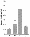
Concentrations of IL-18 in serum and duration of HIV infection. Average ± standard error concentrations of IL-18 in serum of the three groups of patients with different durations of infection. Note the highest serum concentration in the group that has been infected for 3 to 6 years. A, HIV-seronegative control donors; B, C and D, HIV-infected donors with a duration of <3 years, 3 to 6 years, and >6 years of HIV infection, respectively.
FIG. 3.
FIG. 3.
Production of IL-18 from PBMC. PBMC (2 × 10⁵) from HIV-infected/AIDS patients and HIV-seronegative control subjects were cultured in vitro with and without the presence of LPS, and their culture supernatants were collected at the indicated time points and assayed for IL-18 with a commercial ELISA kit. Average ± standard error production of IL-18 in PBMC microculture supernatants (S/N) from eight HIV-infected/AIDS patients (P) and seven control subjects (N) with (A) and without (B) LPS stimulation. At all the time points tested, the patients' PBMC produced less IL-18 than that produced by the control subjects' PBMC.
FIG. 4.
FIG. 4.
Correlation between concentrations of IL-18 in serum and its production from PBMC. (A) Correlation between concentrations of IL-18 in serum and its production from PBMC culture supernatants (S/N) from seven HIV-seronegative control subjects after 12 h of incubation. (B) Correlation between these two parameters from eight HIV-infected/AIDS patients. Similar correlation results were obtained when PBMC were stimulated with LPS.
FIG. 5.
FIG. 5.
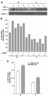
Expression of IL-18 in PBMC. The expression of IL-18 in PBMC was determined by Western blotting using an IL-18-specific monoclonal antibody in 75 μg of cellular lysate. (A) Western blot showing the precursor form of IL-18 (24 kDa) in PBMC with (+) and without (−) stimulation with LPS in HIV-infected/AIDS patients (P) and HIV-seronegative healthy control (N) subjects. The mature form of IL-18 (18 kDa) could not be detected on the blots. (B) Densitometric analysis of the blot showing expression of IL-18 after normalization with β-actin. The 1, 2, and 3 in panels A and B represent data from different donors. (C) Average ± standard error expression of IL-18 in the PBMC.
FIG. 6.
FIG. 6.
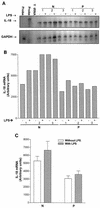
Expression of the IL-18 gene in PBMC. The expression of IL-18 gene in PBMC was determined by RPA with α-32P-labeled cRNA and normalized with respect to GAPDH gene expression (also determined by RPA). (A) Autoradiograph of the polyacrylamide gel showing protected IL-18 and GAPDH-specific probes in the PBMC of HIV-infected/AIDS patients and control subjects with (+) and without (−) stimulation with LPS. Probe and Y tRNA, migration of the labeled cRNA without incubation with mRNA and protection of yeast tRNA (negative control), respectively. (B) Densitometric analysis of the protected probes. Ratio of protected IL-18 and GAPDH probes for each individual. The numbers 1, 2, and 3 in panels A and B represent data for different donors. (C) Average ± standard error expression of the ratio of IL-18 and GAPDH-specific protected probes (IL-18 gene expression). N and P, HIV-seronegative and HIV-infected donors, respectively.
FIG. 7.
FIG. 7.
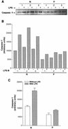
Expression of activated caspase-1 in PBMC. The expression of activated caspase-1 was determined by Western blotting in 75 μg of protein from cell lysates by using an activated caspase-1-specific monoclonal antibody. (A) Western blot showing caspase-1 expression in the PBMC of control HIV-seronegative healthy donors (N) and HIV-infected/AIDS patients (P) with (+) and without (−) stimulation with LPS. (B) Densitometric analysis of the blot. The numbers 1, 2, and 3 in panels A and B represent data from different donors. (C) Average ± standard error expression of caspase-1 in these subjects.
FIG. 8.
FIG. 8.
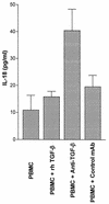
Effect of TGF-β on IL-18 production from PBMC. PBMC (2 × 10⁵) from HIV-seronegative donors were cultured in 200 μl of the culture medium in 96-well plate alone or in the presence of recombinant human (rh) TGF-β (20 ng per ml; R & D Systems), TGF-β-neutralizing, or control antibody (5 μg per ml each; R & D Systems). Culture supernatants were collected 24 h later and assayed for IL-18 content with the ELISA kit. The figure shows average ± standard error IL-18 contents from three replicate wells. Only the addition of the neutralizing antibody resulted in a significant (P < 0.05) increase in TGF-β production.
FIG. 9.
FIG. 9.
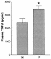
TGF-β contents of the plasma samples. The level of TGF-β was measured in the samples with a commercial ELISA kit. The figure depicts average ± standard error concentration of this cytokine in the samples after its activation (total TGF-β). N and P, HIV-seronegative healthy and HIV-infected donors, respectively. The star on the top of a bar indicates a significant (P < 0.05) difference between the two group means.
FIG. 10.
FIG. 10.
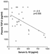
Correlation between the levels of TGF-β in plasma and IL-18 in serum of HIV-infected individuals. The figure depicts a significant negative correlation between these two parameters as determined by the Pearson's method.

Similar articles

Cited by

References

    1. Ahmad, A., R. Ahmad, E. Toma, R. Morisset, and J. Menezes. 2000. Impaired induction of IL-15 in response to herpes simplex virus type 1 infection in peripheral blood mononuclear cells of HIV-infected patients. AIDS 14:744-746. - PubMed
    1. Ahmad, A., E. Sharif-Askari, L. Fawaz, and J. Menezes. 2000. Innate immune response of the human host to exposure with herpes simplex virus type 1: in vitro control of the virus infection by enhanced natural killer activity via interleukin-15 induction. J. Virol. 74:7196-7203. - PMC - PubMed
    1. Ahmad, R., L. Knafo, J. Xu, S. T. Sindhu, J. Menezes, and A. Ahmad. 2000. Thrombin induces apoptosis in human tumor cells. Int. J. Cancer 87:707-715. - PubMed
    1. Akira, S. 2000. The role of IL-18 in innate immunity. Curr. Opin. Immunol. 12:59-63. - PubMed
    1. Allen, J. B., H. L. Wong, P. M. Guyre, G. L. Simon, and S. M. Wahl. 1991. Association of circulating receptor Fc gamma RIII-positive monocytes in AIDS patients with elevated levels of transforming growth factor-beta. J. Clin. Investig. 87:1773-1779. - PMC - PubMed

Publication types

MeSH terms