. 2002 Dec;76(24):12473-82.
doi: 10.1128/jvi.76.24.12473-12482.2002.

African swine fever virus polyproteins pp220 and pp62 assemble into the core shell

Affiliations

African swine fever virus polyproteins pp220 and pp62 assemble into the core shell

Germán Andrés et al. J Virol. 2002 Dec.

Abstract

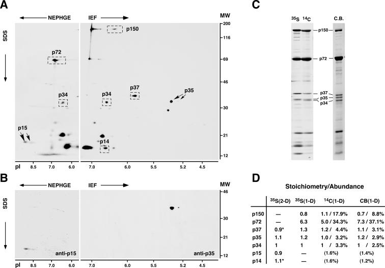
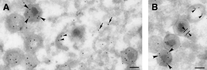
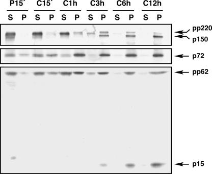
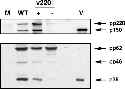
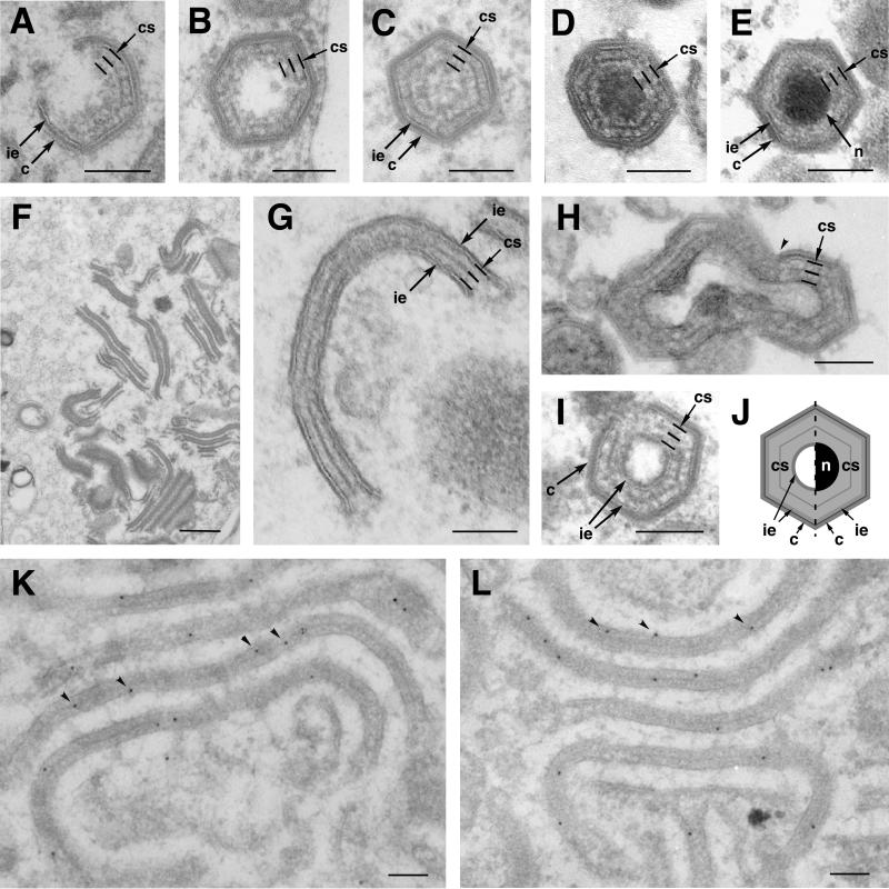
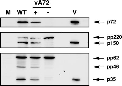
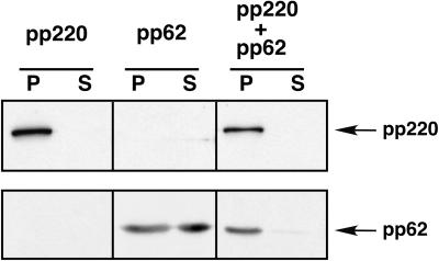
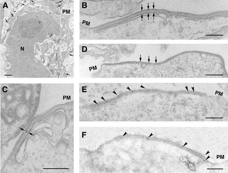
African swine fever virus (ASFV), a complex enveloped DNA virus, expresses two polyprotein precursors, pp220 and pp62, which after proteolytic processing give rise to several major components of the virus particle. We have analyzed the structural role of polyprotein pp62, the precursor form of mature products p35 and p15, in virus morphogenesis. Densitometric analysis of one- and two-dimensional gels of purified virions showed that proteins p35 and p15, as well as the pp220-derived products, are present in equimolecular amounts in the virus particle. Immunoelectron microscopy revealed that the pp62-derived products localize at the core shell, a matrix-like domain placed between the DNA-containing nucleoid and the inner envelope, where the pp220-derived products are also localized. Pulse-chase experiments indicated that the processing of both polyprotein precursors is concomitant with virus assembly. Furthermore, using inducible ASFV recombinants, we show that pp62 processing requires the expression of the pp220 core precursor, whereas the processing of both precursors pp220 and pp62 is dependent on expression of the major capsid protein p72. Interestingly, when p72 expression is blocked, unprocessed pp220 and pp62 polyproteins assemble into aberrant zipper-like elements consisting of an elongated membrane-bound protein structure reminiscent of the core shell. Moreover, the two polyproteins, when coexpressed in COS cells, interact with each other to form zipper-like structures. Together, these findings indicate that the mature products derived from both polyproteins, which collectively account for about 30% of the virion protein mass, are the basic components of the core shell and that polyprotein processing represents a maturational process related to ASFV morphogenesis.


Figures

FIG. 1.
FIG. 1.
Gel mapping, stoichiometry, and relative abundances of ASFV polyprotein products in the virus particle. (A) 2-D gel electrophoresis of ASFV labeled with [35S]methionine. Basic proteins were resolved by NEPHGE, while acidic proteins were separated by IEF. The positions of the major capsid protein p72; the polyprotein pp220-derived products p150, p37, p34, and p14; and the polyprotein pp62-derived products p35 and p15 are indicated. The migrations of molecular weight (MW) markers (in thousands) and of pI markers are indicated at the right and at the bottom of the 2-D gels, respectively. (B) 2-D mapping of proteins p35 and p15. Protein p15 was mapped in NEPHGE gels by immunoprecipitation, whereas p35 was identified in IEF gels by immunoblotting. (C) 1-D gel electrophoresis of ASFV particles labeled with [35S]methionine or 14C-amino acids or stained with Coomassie blue (C.B.). The positions of several structural proteins are indicated. (D) Quantitative analysis of the polyprotein products. Polyprotein products and, as a reference, capsid protein p72 were quantified by densitometry analysis of 2-D gels of [35S]methionine-labeled proteins (two experiments) or of 1-D gels of ASFV proteins labeled with [35S]methionine (three experiments) or 14C-amino acids (two experiments) or stained with Coomassie blue (three experiments). Stoichiometry data were normalized by the number of methionine residues of each protein (for 35S-labeled proteins) or by its molecular weight (for 14C-labeled or Coomassie blue-stained proteins) and referred to protein p34. Relative abundances (percentages) were estimated from 1-D gels of 14C-labeled or Coomassie blue-stained proteins. Standard deviations of the means were less than 25% of the means. Proteins p150 and p72 were not quantified in 2-D gels due to deficient focusing, while proteins p15 and p14 were not estimated in 1-D gels because of heterogeneity of the bands. The relative abundances of proteins p15 and p14 are values extrapolated by assuming an equimolar stoichiometry. Stoichiometry data for p37 and p14 [marked with asterisks in the 35S(2-D) column] are from reference .
FIG. 2.
FIG. 2.
Proteins p35 and p15 reside at the core shell. Thawed cryosections of BA71V-infected cells fixed at 20 hpi were incubated with anti-p35 (A) or anti-p15 (B) antibodies followed by protein A-gold (10-nm diameter). Within the virus factory, the pp62 labeling can be observed close to precursor viral membranes (arrows), at the immature core region of assembling virions (small arrowheads), and at the core shell (large arrowheads) surrounding the electron-dense nucleoid of mature particles. The core shell appears delimited in a virus particle in panel B. Bars, 100 nm.
FIG. 3.
FIG. 3.
Processing of ASFV polyproteins occurs after their incorporation into the membrane-particulate fraction. Infected Vero cells were pulse-labeled with [35S]methionine-[35S]cysteine for 15 min (P15′ lanes) at 12 hpi and either immediately processed or subsequently chased for 15 min, 1 h, 3 h, 6 h, and 12 h (C15′, C1 h, C3 h, C6 h, and C12 h lanes, respectively). The postnuclear supernatants were fractionated into membrane-particulate (lanes P) and soluble (lanes S) fractions and immunoprecipitated with anti-pp220/p150, anti-pp62/p15, and anti-p72 antibodies. The proteins detected in the pulse and chases after SDS-polyacrylamide gel electrophoresis are indicated.
FIG. 4.
FIG. 4.
Polyprotein pp62 processing depends on polyprotein pp220 expression. Vero cells were mock infected (lane M) or infected with parental BA71V (lane WT) or recombinant v220i virus in the presence (lane +) or absence (lane −) of IPTG (isopropyl-β-d-thiogalactopyranoside). At 20 hpi, the cells were lysed and analyzed, together with purified ASFV particles (lane V), by Western immunoblotting with anti-pp220/p150 and anti-pp62/p35 antibodies. The positions of polyprotein pp220 and its derived protein p150 (upper panel) and of polyprotein pp62, the intermediate processing precursor pp46, and the mature product p35 (lower panel), are indicated.
FIG. 5.
FIG. 5.
The core shell and the aberrant zipper-like structures are structurally and biochemically related. Cells infected with parental BA71V (A, B, C, D, and E) and recombinant vA72 (F, G, H, and I) were processed by conventional Epon embedding at 16 to 24 hpi. (A to E) Different stages of the assembly of an intracellular mature particle. Note that the assembly of the core shell (indicated by three parallel lines) beneath the inner envelope is concomitant with the formation of the outer capsid (A) and precedes the maturation of the DNA-containing nucleoid (B to D). Note also that the fine electron-dense layer that subdivides the core shell is less evident in intracellular mature virions (E). (F and G) Aberrant zipper-like structures assembled at vA72 virus factories at 16 hpi under restrictive conditions. Zipper-like elements consist of a core shell-like structure limited by two inner envelopes (G). (H and I) Icosahedral aberrant viruses formed in vA72-infected cells maintained for 16 h under restrictive conditions and then treated with inducer for an additional 8-h period. Note that the zipper elements become icosahedral, double-enveloped particles after capsid assembly (arrowhead in panel H) on one of the two flanking envelopes. Note also that the core shell of these particles (I) is nearly identical to that of normal assembling virions (B to D). (J) A diagram illustrates the similarities and differences between a normal (right half) and an aberrant (left half) particle. Note that the aberrant particle contains an additional innermost lipid envelope but lacks the nucleoid. (K and L) Immunogold labeling of vA72-infected cells maintained under restrictive conditions for 24 h. Lowicryl sections were labeled with anti-pp62 (K) or anti-pp220 (L) antibodies followed by protein A-gold (10-nm diameter). c, capsid; ie, inner envelope; cs, core shell; n, nucleoid. Bars, 100 nm.
FIG. 6.
FIG. 6.
Proteolytic processing of core polyproteins pp220 and pp62 requires the expression of the major capsid protein p72. Vero cells were mock infected (lane M) or infected for 20 h with parental BA71V (lane WT) or recombinant vA72 virus in the presence (lane +) or absence (lane −) of IPTG. Cell extracts were analyzed, together with purified ASFV particles (lane V), by immunoblotting with antibodies against capsid protein p72, polyprotein pp220 and mature product p150, or polyprotein pp62 and mature product p35. The positions of the detected proteins are indicated.
FIG. 7.
FIG. 7.
Coexpression of polyproteins pp220 and pp62 in COS-7 cells. COS-7 cells were transfected with plasmids bearing the pp220- or pp62-encoding genes or cotransfected with both plasmids and infected with vaccinia virus vTF7-3 in the presence of Ara C. At 7 hpi, the cells were fractionated into a low-speed sediment (lanes P) and a postnuclear supernatant (lanes S) and the fractions were analyzed by Western immunoblotting with anti-pp220 or anti-pp62 serum.
FIG. 8.
FIG. 8.
Polyproteins pp220 and pp62 assemble into zipper-like structures. COS-7 cells were cotransfected with plasmids bearing the pp220- and pp62-encoding genes and infected with vaccinia virus vTF7-3 in the presence of Ara C. At 12 hpi, the cells were processed for conventional Epon embedding (B, C, and D) or freeze-substitution and Lowicryl embedding (A, E, and F). (A) A cotransfected cell containing pp220- and pp62-induced zipper-like structures in different areas of the cell surface (arrows). (B and C) At a higher magnification, the zipper-like structures reveal a symmetrical organization consisting of a trilaminar cytosolic domain of about 32 nm limited by lipid membranes (arrows). Two thick layers separated by a thin electron-dense layer (line in panel C) compose the protein domain. (D) For comparison, a pp220-induced coat, which is bound to only one membrane and is approximately 24 nm thick, is shown. (E and F) The Lowicryl sections of cotransfected cells were incubated with anti-pp220 (E) or anti-pp62 (F) serum followed by protein A-gold (10-nm diameter). Note that both antibodies label zipper-like structures associated with the plasma membrane (PM) and intracellular cisternae. Bars, 100 nm (A) and 200 nm (B to F).

References

    1. Alonso, C., J. Miskin, B. Hernáez, P. Fernández-Zapatero, L. Soto, C. Cantó, I. Rodríguez-Crespo, L. Dixon, and J. M. Escribano. 2001. African swine fever virus protein p54 interacts with the microtubular motor complex through direct binding to light-chain dynein. J. Virol. 75:9819-9827. - PMC - PubMed
    1. Alves de Matos, A. P., and Z. G. Carvalho. 1993. African swine fever virus interaction with microtubules. Biol. Cell 78:229-234. - PubMed
    1. Andrés, G., C. Simón-Mateo, and E. Viñuela. 1993. Characterization of two African swine fever virus 220-kDa proteins: a precursor of the major structural protein p150 and an oligomer of phosphoprotein p32. Virology 194:284-293. - PubMed
    1. Andrés, G., C. Simón-Mateo, and E. Viñuela. 1997. Assembly of African swine fever virus: role of polyprotein pp220. J. Virol. 71:2331-2341. - PMC - PubMed
    1. Andrés, G., R. García-Escudero, C. Simón-Mateo, and E. Viñuela. 1998. African swine fever virus is enveloped by a two-membraned collapsed cisterna derived from the endoplasmic reticulum. J. Virol. 72:8988-9001. - PMC - PubMed

Publication types