. 2002 Dec;76(24):12491-502.
doi: 10.1128/jvi.76.24.12491-12502.2002.

Coronaviruses maintain viability despite dramatic rearrangements of the strictly conserved genome organization

Affiliations

Coronaviruses maintain viability despite dramatic rearrangements of the strictly conserved genome organization

Cornelis A M de Haan et al. J Virol. 2002 Dec.

Abstract

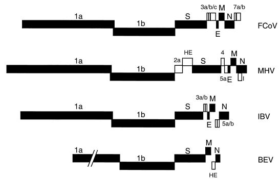
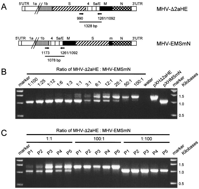
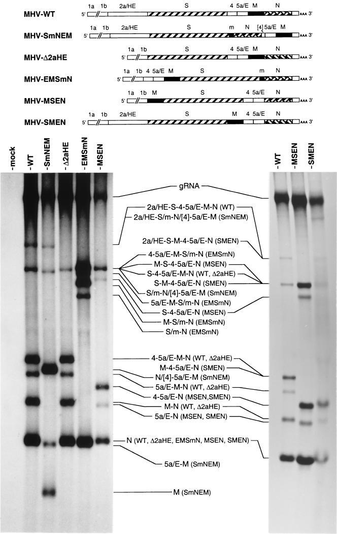
Despite their high frequency of RNA recombination, the plus-strand coronaviruses have a characteristic, strictly conserved genome organization with the essential genes occurring in the order 5'-polymerase (pol)-S-E-M-N-3'. We have investigated the significance of this remarkable conservation by rearrangement of the murine coronavirus genome through targeted recombination. Thus, viruses were prepared with the following gene order: 5'-pol-S-M-E-N-3', 5'-pol-S-N-E-M-3', 5'-pol-M-S-E-N-3', and 5'-pol-E-M-S-N-3'. All of these viruses were surprisingly viable, and most viruses replicated in cell culture with growth characteristics similar to those of the parental virus. The recombinant virus with the gene order 5'-pol-E-M-S-N-3' was also tested for the ability to replicate in the natural host, the mouse. The results indicate that the canonical coronavirus genome organization is not essential for replication in vitro and in vivo. Deliberate rearrangement of the viral genes may be useful in the generation of attenuated coronaviruses, which due to their reduced risk of generating viable viruses by recombination with circulating field viruses, would make safer vaccines.


Figures

FIG. 1.
FIG. 1.
Genomic organization of the Coronaviridae. A member of each coronavirus subgroup—the group I feline coronavirus (FcoV), group II mouse hepatitis virus (MHV), and group III infectious bronchitis virus (IBV)—and the Berne torovirus (BEV) are represented. The essential genes are represented by solid boxes, while the (presumed) nonessential genes are represented by open boxes.
FIG. 2.
FIG. 2.
Plasmid constructs, targeted recombination, and recombinant viruses. The transcription vectors from which the defective RNAs were produced in vitro by using T7 RNA replicase are indicated on the left. Vector pMH54 has been described before (17). The other vectors were derived from pMH54 as described in Materials and Methods. The arrow at the left end of each vector indicates the T7 promoter; the solid circle represents the polylinker between the 5′-end segment of the MHV genome (labeled 5′/1) and either the HE gene or the 3′ end of the replicase gene (1b), followed by the structural and group-specific genes and the 3′ untranslated region (UTR) and the polyadenylated segment (together labeled 3′/A). “m” indicates a 126-nt segment from the 3′end of the M gene. The positions of the sequences shown in Fig. 3 are indicated by arrowheads (the numbers correspond to those in the circles in Fig. 3). On top, a scheme for targeted recombination using the interspecies chimeric virus fMHV, which grows only in feline cells, is shown. Recombinant viruses generated by the indicated crossover event can be selected on the basis of their ability to grow in murine cells. The genomes of these viruses are represented on the right.
FIG. 3.
FIG. 3.
Sequences of the numbered gene junctions shown in Fig. 2. The numbers in the circles correspond to those under the arrowheads in Fig. 2. Nucleotides that differ from those in the original wild-type MHV A59 sequence are underlined and in boldface.
FIG. 4.
FIG. 4.
Single-step growth kinetics of MHV recombinants. LR7 cells were infected with each recombinant MHV at an MOI of 8 PFU per cell. Viral infectivities in culture media at different times postinfection were determined by a quantal assay on LR7 cells, and the TCID50 were calculated.
FIG. 5.
FIG. 5.
Relative fitness of MHV-Δ2aHE and MHV-EMSmN in cell culture. (A) competitive RT-PCR was used to evaluate the relative yields of MHV-Δ2aHE and MHV-EMSmN after each passage. Primers used in the experiment, their approximate loci in the MHV genome, and the predicted sizes of the PCR products are indicated. Primer 1092 was used in the RT step, while primers 1261, 990, and 1173 were used for the PCR (3). PCR products were analyzed by electrophoresis in 1% agarose gels stained with ethidium bromide. (B) As a reference, MHV-Δ2aHE and MHV-EMSmN were mixed in different ratios, and RT-PCR was performed on purified genomic RNA. As controls, PCR products obtained with plasmids used for the construction of the recombinant viruses and with water are shown. (C) MHV-Δ2aHE and MHV-EMSmN were mixed in 1:100, 1:1, and 100:1 infectivity ratios and inoculated onto LR7 cells. The culture supernatant harvested after 16 h (P1) was passaged four additional times at a low MOI on the same cells (P2 to P5). RT-PCR was performed on genomic RNA purified from aliquots of the successive culture media, and the products were analyzed again in 1% agarose gels. The sizes of relevant DNA fragments of the marker are indicated to the right of each gel.
FIG.6.
FIG.6.
Analysis of intracellular RNA synthesis by recombinant MHVs with rearranged genomes. Mouse 17Cl1 cells were mock infected (mock) or infected with recombinant virus (MHV-WT, MHV-SmNEM, MHV-Δ2aHE, MHV-EMSmN, or MHV-MSEN) at an MOI of 5 PFU per cell and were labeled from 7 to 9 h postinfection with [33P]orthophosphate as described in Materials and Methods. In the case of MHV-SMEN, where the maximal MOI that could be achieved was approximately 0.02 PFU per cell, labeling was carried out from 24 to 26 h postinfection. Purified total cytoplasmic RNA was separated by electrophoresis through 1% agarose containing formaldehyde, and the labeled RNA was visualized by fluorography. The genomic composition of each virus is indicated schematically at the top, and each RNA species, with its viral sources in parentheses, is indicated between the panels. Note that in the right-hand gel, the RNA sample in the MHV-SMEN lane was from five times as many cells as that in the MHV-WT lane.
FIG. 7.
FIG. 7.
Analysis of synthesis of recombinant MHV proteins. (A) LR7 cells were mock infected (mock) or infected with recombinant viruses as indicated on the top of the gel. The cells were labeled for 2 h with 35S-labeled amino acids starting 6 h postinfection. Total lysates were prepared and used for immunoprecipitation with the anti-MHV serum K134, and the precipitates were analyzed by SDS-15% PAGE. The positions of the different proteins are indicated on the left, while the molecular mass marker is indicated on the right of the gel. (B) For quantitative analysis, PhosphorImager scanning of the amounts of radioactivity in the M, S, and N proteins in the dried gels from three independent experiments (as shown in panel A) was performed. The ratios of the amounts of M and N (M/N) and S and N (S/N) proteins were calculated relative to those of MHV-WT-infected cells, and standard deviations are indicated.
FIG. 8.
FIG. 8.
Replication of recombinant viruses in BALB/c mice. Eight-week-old mice were inoculated intraperitoneally with a 100-μl volume containing 106 TCID50 of recombinant virus (n = 4 per virus). The mice were sacrificed, and the livers were removed on day 4 postinfection. Virus titers were determined by plaque assay on LR7 cell monolayers following homogenization of the livers. The error bars indicate standard deviations.

References

    1. Ball, L. A., C. R. Pringle, B. Flanagan, V. P. Perepelitsa, and G. W. Wertz. 1999. Phenotypic consequences of rearranging the P, M, and G genes of vesicular stomatitis virus. J. Virol. 73:4705-4712. - PMC - PubMed
    1. de Haan, C. A. M., L. Kuo, P. S. Masters, H. Vennema, and P. J. M. Rottier. 1998. Coronavirus particle assembly: primary structure requirements of the membrane protein. J. Virol. 72:6838-6850. - PMC - PubMed
    1. de Haan, C. A. M., P. S. Masters, X. Shen, S. Weiss, and P. J. M. Rottier. 2002. The group-specific murine coronavirus genes are not essential, but their deletion, by reverse genetics, is attenuating in the natural host. Virology 296:177-189. - PMC - PubMed
    1. de Vries, A. A. F., A. L. Glaser, M. J. Raamsman, C. A. M. de Haan, S. Sarnataro, G. J. Godeke, and P. J. M. Rottier. 2000. Genetic manipulation of equine arteritis virus using full-length cDNA clones: separation of overlapping genes and expression of a foreign epitope. Virology 270:84-97. - PubMed
    1. de Vries, A. A. F., A. L. Glaser, M. J. Raamsman, and P. J. M. Rottier. 2001. Recombinant equine arteritis virus as an expression vector. Virology 284:259-276. - PubMed

Publication types

LinkOut - more resources