. 2002 Dec;76(24):12813-22.
doi: 10.1128/jvi.76.24.12813-12822.2002.

The complementary strand of the human T-cell leukemia virus type 1 RNA genome encodes a bZIP transcription factor that down-regulates viral transcription

Affiliations

The complementary strand of the human T-cell leukemia virus type 1 RNA genome encodes a bZIP transcription factor that down-regulates viral transcription

Gilles Gaudray et al. J Virol. 2002 Dec.

Abstract

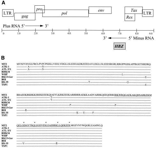
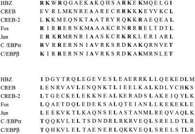
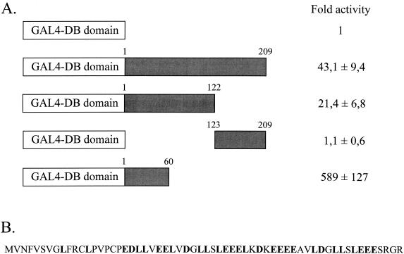
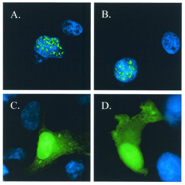
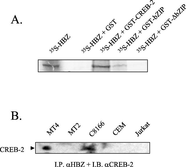
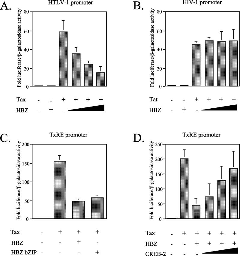
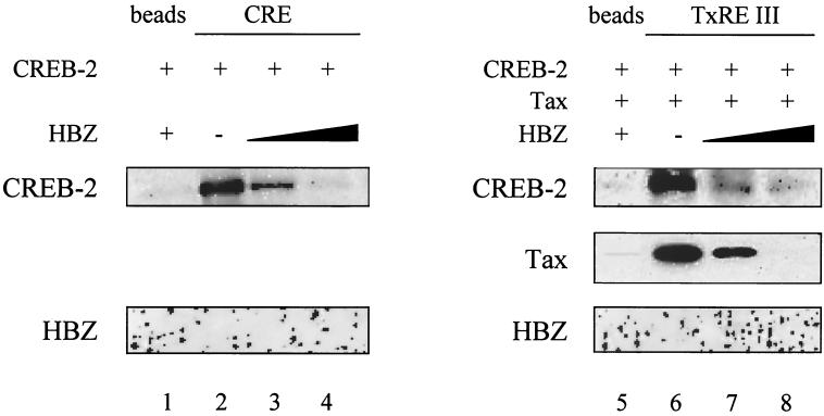
The RNA genome of the human T-cell leukemia virus type 1 (HTLV-1) codes for proteins involved in infectivity, replication, and transformation. We report in this study the characterization of a novel viral protein encoded by the complementary strand of the HTLV-1 RNA genome. This protein, designated HBZ (for HTLV-1 bZIP factor), contains a N-terminal transcriptional activation domain and a leucine zipper motif in its C terminus. We show here that HBZ is able to interact with the bZIP transcription factor CREB-2 (also called ATF-4), known to activate the HTLV-1 transcription by recruiting the viral trans-activator Tax on the Tax-responsive elements (TxREs). However, we demonstrate that the HBZ/CREB-2 heterodimers are no more able to bind to the TxRE and cyclic AMP response element sites. Taking these findings together, the functional inactivation of CREB-2 by HBZ is suggested to contribute to regulation of the HTLV-1 transcription. Moreover, the characterization of a minus-strand gene protein encoded by HTLV-1 has never been reported until now.


Figures

FIG. 1.
FIG. 1.
The HTLV-1 genome encodes the bZIP transcription factor HBZ. (A) Organization of the HTLV-1 genome. The HTLV-1 provirus genome (in kilobase pairs) is represented by a line. The viral genes encoded by the plus-strand RNA (white boxes) are shown above the genome line (only the genes whose functions have been clearly established are represented). The HBZ gene encoded by the minus-strand RNA is represented by a greyish box, below the line. (B) Comparison of the amino acid sequence of HBZ characterized from the HTLV-1-infected MT2 cell line with the HBZ protein from different HTLV-1 isolates. The amino acid sequence of MT2 HBZ is shown. The bZIP domain is underlined, and the leucine residues within the leucine zipper are indicated by asterisks. Only differences in the other HTLV-1 isolates are indicated, while homologies are represented by successive dashes. The different strains (with GenBank accession numbers in parentheses) are ATK-1 (J02029), ATL-YS (U19949), RHK34 (L03562), WHP (AF259264), RK13-Ger (AF042071), BOI (L36905), HS-35 (D13784), and TSP1 (M86840).
FIG. 2.
FIG. 2.
Comparison of the amino acid sequences of the basic domains (at top; residues 140 to 164) and the leucine zipper structures (at bottom; residues 164 to 193) of HBZ and several other bZIP transcription factors. The conserved amino acids are indicated in boldface type.
FIG. 3.
FIG. 3.
The N terminus of HBZ contains a potent transcriptional activation domain. (A) CEM cells were cotransfected with 5 μg of pACβ1 (β-galactosidase containing reference plasmid), 2 μg of the luciferase reporter vector pG5luc, and 2 μg of the eukaryotic vector pBIND expressing the GAL4 DB domain alone or fused to either the full-length HBZ product (209 amino acids long), the N-terminal region (residues 1 to 122 and 1 to 60), or the C-terminal domain (residues 123 to 209). Luciferase values were normalized for β-galactosidase activity and are expressed as increases relative to that of cells transfected with pSG-5 and pBIND only expressing the GAL4 DB domain. Values are presented as means ± standard deviations (n = 3). The GAL4-DB domain and the different regions of HBZ are represented by white and greyish boxes, respectively. (B) Amino acid sequence of the HBZ N terminus (residues 1 to 60). The N terminus is rich in acidic and leucine residues (indicated in boldface type in the sequence).
FIG. 4.
FIG. 4.
Detection of HBZ in vivo by immunoprecipitation. Proteins were immunoprecipitated from the lysate of either the HTLV-1-infected T-cell line C8166 or the uninfected T-cell line CEM using preimmune sera (PI) or the anti-HBZ sera (I) produced by two different rabbits (A and B). Precipitates were analyzed by SDS-PAGE and immunoblotting with anti-HBZ serum. The molecular mass markers (in kilodaltons) are indicated on the right of the figure. (C) The same approach was carried out from the lysate of the HTLV-1-infected MT4 cell line using preimmune serum (PI) or the anti-HBZ serum (I).
FIG. 5.
FIG. 5.
Immunofluorescence microscopy analysis of the subcellular localization of HBZ with an N-terminal GFP tag in vivo. COS7 cells were transfected with expression vectors encoding either GFP-HBZ (A and B), GFP (C), or GFP-HBZΔbZIP (D). Cells were cultivated on the glass slides, fixed, and stained with Hoechst solution, and then the green fluorescence was analyzed by immunofluorescence microscopy. The specificity of the immunofluorescent staining is indicated by the absence of signal in flanking untransfected cells. The blue fluorescence of the nuclei (Hoechst staining) is visualized by UV illumination.
FIG. 6.
FIG. 6.
HBZ interacts with CREB-2. (A) In vitro binding assays of HBZ to CREB-2. Equal amounts of GST, GST-CREB-2, GST-CREB-2263-351 (lane GST-bZIP), and GST-CREB-21-262 (lane GST-ΔbZIP) immobilized on glutathione-Sepharose beads were incubated with [35S]HBZ, and bound proteins were analyzed by SDS-PAGE and autoradiography. The first lane corresponds to in vitro-translated [35S]HBZ. (B) Binding of HBZ to CREB-2 in vivo. Proteins from MT4, MT2, C8166, CEM, and Jurkat cell total lysate were immunoprecipitated with rabbit anti-HBZ (I.P. αHBZ), and immunoprecipitated proteins were analyzed by immunoblotting with goat anti-CREB-2 (I.B. αCREB-2).
FIG. 7.
FIG. 7.
HBZ down-regulates the HTLV-1 transcription by interacting with CREB-2. CEM cells were cotransfected with the following: (A) 2 μg of HTLV-1 LTR-luciferase, 1 μg of Tax expression vector pSG-Tax, and pCI-HBZ (0, 1, 3, or 9 μg) (the luciferase values are expressed as increases relative to that of cells transfected with pSG-5, pCI-neo, and HTLV-1 LTR-luciferase); (B) 2 μg of HIV-1 LTR-luciferase, 1 μg of the Tat expression vector pBg312HIV-1Lai-Tat, and pCI-HBZ (0, 1, 3, or 9 μg) (the luciferase values are expressed as increases relative to that of cells transfected with HIV-1 LTR-luciferase without Tat and HBZ); (C) 2 μg of HTLV-1 TxRE-luciferase, 1 μg of pSG-Tax, and 5 μg of pCI-neo expressing either HBZ or the HBZ bZIP domain (the luciferase values are expressed as increases relative to that of cells transfected with pSG-5, pCI-neo, and HTLV-1 TxRE-luciferase); (D) 2 μg of HTLV-1 TxRE-luciferase, 1 μg of pSG-Tax, 5 μg of pCI-HBZ, and pCI-CREB-2 (0, 1, 3, or 9 μg) (the luciferase values are expressed as fold increase relative to that of cells transfected with pSG-5, pCI-neo, and HTLV-1 TxRE-luciferase). For all the cotransfections, the total amount of DNA in each series of transfection was equal, the balance being made up with empty plasmids. and the luciferase values were normalized for β-galactosidase activity. Values represent means plus standard deviations (error bars) (n = 3).
FIG. 8.
FIG. 8.
HBZ abolishes the binding of CREB-2 to the CRE and TxRE sites. Biotinylated oligonucleotides (100 ng) corresponding to the somatostatin CRE were incubated with 50 ng of CREB-2 in the absence (lane 2) or the presence (lanes 3 and 4) of the bZIP domain of HBZ (25 and 50 ng). The same experiment was performed with the HTLV-1 TxRE III, CREB-2, and Tax (50 ng) in the absence (lane 6) or the presence (lanes 7 and 8) of HBZ bZIP.

References

    1. Anderson, M. G., and W. S. Dynan. 1994. Quantitative studies of the effect of HTLV-I Tax protein on CREB protein-DNA binding. Nucleic Acids Res. 22:3194-3201. - PMC - PubMed
    1. Cibelli, G., S. Schoch, and G. Thiel. 1999. Nuclear targeting of cAMP response element binding protein 2 (CREB2). Eur. J. Cell Biol. 78:642-649. - PubMed
    1. Coudronniere, N., J. Corbeil, V. Robert-Hebmann, J. M. Mesnard, and C. Devaux. 1998. The lck protein tyrosine kinase is not involved in antibody-mediated CD4 (CDR3-loop) signal transduction that inhibits HIV-1 transcription. Eur. J. Immunol. 28:1445-1457. - PubMed
    1. Franklin, A. A., M. F. Kubik, M. N. Uittenbogaard, A. Brauweiler, P. Utaisincharoen, M. A. Matthews, W. S. Dynan, J. P. Hoeffler, and J. K. Nyborg. 1993. Transactivation by the human T-cell leukemia virus Tax protein is mediated through enhanced binding of activating transcription factor-2 (ATF-2) ATF-2 response and cAMP element-binding protein (CREB). J. Biol. Chem. 268:21225-21231. - PubMed
    1. Franklin, A. A., and J. K. Nyborg. 1995. Mechanisms of Tax regulation of human T cell leukemia virus type I gene expression. J. Biomed. Sci. 2:17-29. - PubMed

Publication types

MeSH terms

LinkOut - more resources