

doi: 10.1128/jvi.76.24.13097-13100.2002.
Identification of neutralizing epitopes within structural domain III of the West Nile virus envelope protein
Affiliations
- PMID: 12438639
- PMCID: PMC136710
- DOI: 10.1128/jvi.76.24.13097-13100.2002
Item in Clipboard
Identification of neutralizing epitopes within structural domain III of the West Nile virus envelope protein
J Virol.
2002 Dec.
Abstract
Using a panel of neutralizing monoclonal antibodies, we have mapped epitopes in domain III of the envelope protein of the New York strain of West Nile virus. The ability of monoclonal antibodies that recognize these epitopes to neutralize virus appeared to differ between lineage I and II West Nile virus strains, and epitopes were located on the upper surface of domain III at residues E307, E330, and E332.
Figures
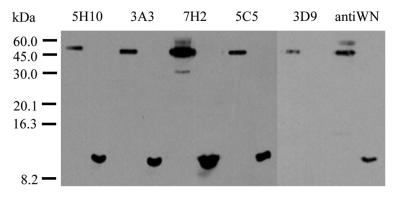
Reactions of anti-WN virus E protein MAbs and polyclonal mouse hyperimmune ascitic fluid (“antiWN”) with viral E protein in a WN virus-infected Vero cell lysate (left lane of each set) or purified, recombinant WN virus E protein domain III (right lane of each set) in a nonreducing Western blot.
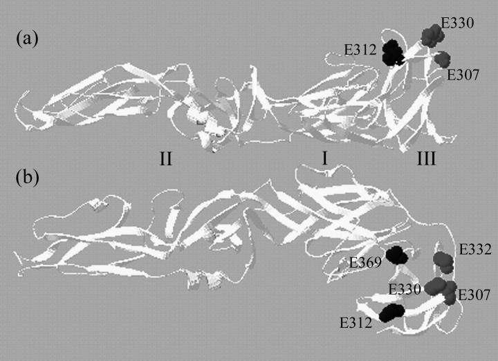
Locations of residues (E307, E330, and E332, shown in dark gray) associated with escape from neutralization by MAbs and/or binding to mouse brain tissue MRP in side-on (a) and overhead (b) views of a predicted structure of the WN virus E protein based on that of the tick-borne encephalitis virus E protein crystal structure (13). Other domain III residues that differed between strains 385-99 and H-442 (E312 and E369) are shown in black. The WN virus structure was derived with the Swiss-Model structure prediction server via Expasy (www.expasy.ch ) and drawn with Swiss-PDB Viewer (7).
References
-
- Beasley, D. W. C., L. Li, M. T. Suderman, and A. D. T. Barrett. 2001. West Nile virus strains differ in mouse neurovirulence and binding to mouse or human brain membrane receptor preparations. Ann. N. Y. Acad. Sci. 951:332-335. - PubMed
-
- Beasley, D. W. C., L. Li, M. T. Suderman, and A. D. T. Barrett. 2002. Mouse neuroinvasive phenotype of West Nile virus strains varies depending upon virus genotype. Virology 296:17-23. - PubMed
-
- Cecilia, D., and E. A. Gould. 1991. Nucleotide changes responsible for loss of neuroinvasiveness in Japanese encephalitis virus neutralization-resistant mutants. Virology 181:70-77. - PubMed
-
- Chambers, T. J., M. Halevy, A. Nestorowicz, C. M. Rice, and S. Lustig. 1998. West Nile virus envelope proteins: nucleotide sequence analysis of strains differing in mouse neuroinvasiveness. J. Gen. Virol. 79:2375-2380. - PubMed
Publication types
MeSH terms
Substances
LinkOut - more resources
Full Text Sources
Other Literature Sources
Molecular Biology Databases
