Comparative Study
. 2002 Nov 26;99(24):15357-62.
doi: 10.1073/pnas.232517099. Epub 2002 Nov 15.

Structure and function from the circadian clock protein KaiA of Synechococcus elongatus: a potential clock input mechanism

Affiliations
Comparative Study

Structure and function from the circadian clock protein KaiA of Synechococcus elongatus: a potential clock input mechanism

Stanly B Williams et al. Proc Natl Acad Sci U S A. .

Erratum in

  • Proc Natl Acad Sci U S A. 2003 Jan 21;100(2):763.

Abstract

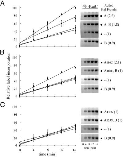
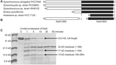
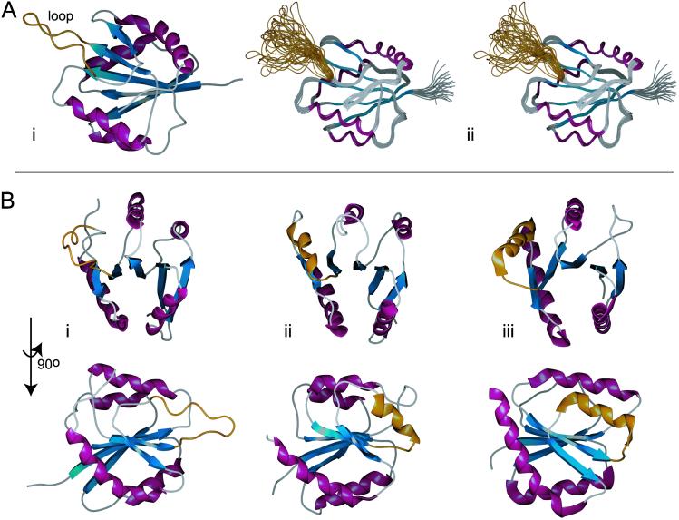
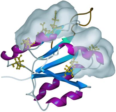
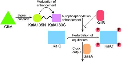
In the cyanobacterium Synechococcus elongatus (PCC 7942) the proteins KaiA, KaiB, and KaiC are required for circadian clock function. We deduced a circadian clock function for KaiA from a combination of biochemical and structural data. Both KaiA and its isolated carboxyl-terminal domain (KaiA180C) stimulated KaiC autophosphorylation and facilitated attenuation of KaiC autophosphorylation by KaiB. An amino-terminal domain (KaiA135N) had no function in the autophosphorylation assay. NMR structure determination showed that KaiA135N is a pseudo-receiver domain. We propose that this pseudo-receiver is a timing input-device that regulates KaiA stimulation of KaiC autophosphorylation, which in turn is essential for circadian timekeeping.


Figures

Fig 1.
Fig 1.
Influence of Kai proteins on the autophosphorylation rate of KaiC. (A) Assays were run under initial rate conditions as described (see Materials and Methods). The graph illustrates the time course of KaiC autophosphorylation with ATP in the presence of KaiA, KaiA and KaiB, no additional protein, and KaiB. All time points are the average of four independent experiments, including the one represented by the gel images. Error bars indicate standard deviation from the mean (n = 4). Relative rates calculated from the line slopes are indicated parenthetically. (B) As above but the KaiA180C domain was substituted for KaiA in each assay. (C) As above but the Kai135N domain was substituted for KaiA in each assay.
Fig 2.
Fig 2.
(A) Graphic representation of five apparent KaiA homologs from the listed cyanobacteria. The amino termini are regions of low sequence similarity and are represented by white boxes. The carboxyl termini are regions of high sequence similarity and are represented by black boxes. (B) Graphic representation of the two independent domains derived from S. elongatus KaiA in this study. (C) An SDS/PAGE gel containing samples from a limited trypsin digest assay (see Materials and Methods). As indicated, we observed three major species. Production of the KaiA135N and KaiA180C domains was, in part, based on the largest and smallest of the three species.
Fig 3.
Fig 3.
The solution structure of KaiA135N and comparisons to other receiver domain proteins. β-strands are in blue, α-helices are in purple, and the flexible loop of KaiA135N and the equivalent region of other receiver domains are in gold. The structure coordinates have been deposited in the Protein Data Bank under PDB ID codes and for the average minimized structure and the family of structures, respectively. (Ai) Schematic representation of the average minimized structure. The solution structure of KaiA135N is an α-β-α sandwich built around a five-parallel-strand β-sheet with b-a-c-d-e arrangement. The rotational correlation time (τc) was calculated to be ≈8.2 ns, which is consistent with a monomer in solution (21). (Aii) Stereoview of the overlaid backbone of a family of 25 low-energy structures calculated from 2,034 distance and geometry restraints. The backbone rms deviation from the average is 0.38 ± 0.04 Å for residues 4–83 and 98–135. The rms deviation for all heavy atoms is 0.78 ± 0.05 Å for the same residues. Few medium- or long-range NOE contacts were identified for residues 83–97, and 15N dynamics (see supporting information) showed that this region is highly dynamic. (B) Structural comparison of KaiA135N with other receiver domains. Shown here are KaiA135N (Bi), the NtrC (1DC7) receiver domain (Bii), and the AmiR (1QO0, residues 11–131) receiver domain (Biii) at two mutually orthogonal views. Figures were prepared with spock (38).
Fig 4.
Fig 4.
Stable KaiA135N clock-period altering mutations and a putative protein-interacting surface. Shown here is the average minimized structure of KaiA135N. Residue substitutions that yield stable proteins in vitro are shown in gold. Most of these residues are mapped on α2 or α4 (I16F, Q113R, Q117L, D119E, D119G). Chemical shift analysis of the mutant proteins versus the wild-type protein showed changes extending over these two helices. Shown here is the water-accessible surface of the residues affected by these mutations. We postulate that this surface is important for signal transduction either between KaiA135N and an upstream signal component or between the two KaiA domains.
Fig 5.
Fig 5.
Working model of KaiA protein function and its role in S. elongatus circadian timekeeping. CikA and other environmental sensors initiate signal transduction cascades that result in activation of the KaiA pseudo-receiver domain. This activation modulates the KaiA carboxyl-terminal domain's enhancement of the KaiC autophosphorylation rate. Thus, equilibria between KaiC phosphorylation states are perturbed. These states differentially control clock output, possibly through the SasA protein kinase. In this manner, a cycle of input, oscillation, and output can be established.

References

    1. Golden S. S., Johnson, C. H. & Kondo, T. (1998) Curr. Opin. Microbiol. 1 669-673. - PubMed
    1. Ishiura M., Kutsuna, S., Aoki, S., Iwasaki, H., Andersson, C. R., Tanabe, A., Golden, S. S., Johnson, C. H. & Kondo, T. (1998) Science 281 1519-1523. - PubMed
    1. Young M. W. & Kay, S. A. (2001) Nat. Rev. Genet. 2 702-715. - PubMed
    1. Lorne J., Scheffer, J., Lee, A., Painter, M. & Miao, V. P. (2000) FEMS Microbiol. Lett. 189 129-133. - PubMed
    1. Iwasaki H., Taniguchi, Y., Ishiura, M. & Kondo, T. (1999) EMBO J. 18 1137-1145. - PMC - PubMed

Publication types

MeSH terms

Substances

LinkOut - more resources