. 2002 Nov 26;99(24):15675-80.
doi: 10.1073/pnas.232568599. Epub 2002 Nov 15.

Killing of Caenorhabditis elegans by Cryptococcus neoformans as a model of yeast pathogenesis

Affiliations

Killing of Caenorhabditis elegans by Cryptococcus neoformans as a model of yeast pathogenesis

Eleftherios Mylonakis et al. Proc Natl Acad Sci U S A. .

Abstract

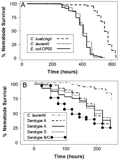
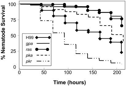
We found that the well-studied nematode Caenorhabditis elegans can use various yeasts, including Cryptococcus laurentii and Cryptococcus kuetzingii, as a sole source of food, producing similar brood sizes compared with growth on its usual laboratory food source Escherichia coli OP50. C. elegans grown on these yeasts had a life span similar to (C. laurentii) or longer than (C. kuetzingii) those fed on E. coli. However, the human pathogenic yeast Cryptococcus neoformans killed C. elegans, and the C. neoformans polysaccharide capsule as well as several C. neoformans genes previously shown to be involved in mammalian virulence were also shown to play a role in C. elegans killing. These included genes associated with signal transduction pathways (GPA1, PKA1, PKR1, and RAS1), laccase production (LAC1), and the alpha mating type. C. neoformans adenine auxotrophs, which are less virulent in mammals, were also less virulent in C. elegans. These results support the model that mammalian pathogenesis of C. neoformans may be a consequence of adaptations that have evolved during the interaction of C. neoformans with environmental predators such as free-living nematodes and amoebae and suggest that C. elegans can be used as a simple model host in which C. neoformans pathogenesis can be readily studied.


Figures

Fig 1.
Fig 1.
(A) Life span of wild-type C. elegans N2 on NGM plates containing 5-fluoro-2′-deoxyuridine (FdUrd) feeding on lawns of C. kuetzingii ATCC#42276, C. laurentii ATCC#18803, and E. coli OP50. P < 0.0001 for E. coli OP50 or C. laurentii compared with C. kuetzingii. Similar results were obtained with C. laurentii strains ATCC#66036 and ATCC#76483. (B) Survival of C. elegans N2 feeding on lawns of C. neoformans serotype A (ATCC#62067, ATCC#62068), serotype B/C (ATCC#34877), or serotype D (ATCC#36556), or on a lawn of C. laurentii ATCC#18803. P < 0.001 for each of the C. neoformans strains compared with C. laurentii.
Fig 2.
Fig 2.
C. neoformans but not C. laurentii accumulates in the gastrointestinal tract of C. elegans. (A) Intact yeast cells present in the distended gastrointestinal tract after feeding 24 h on C. neoformans strain H99. (B) No C. laurentii cells can be detected in the gastrointestinal tract after 24 h of feeding. The round structure (white arrows) is the pharyngeal grinder organ, which functions to disrupt ingested organisms. Black and gray arrows point to the intestinal lumen.
Fig 3.
Fig 3.
C. neoformans virulence factors for mammalian infection also enhance killing of C. elegans. Survival of C. elegans N2 animals feeding on C. neoformans mutants with disruptions in the genes encoding the G protein-cAMP-PKA and the RAS1-controlled signal transduction cascades demonstrated hypovirulence (gpa1, ras1, and pka1) or hypervirulence (pkr1), similar to results in mammalian models. P < 0.001 for each of the mutants compared with the parental strain H99 (see Table 1).
Fig 4.
Fig 4.
The C. neoformans MFα1 promoter is specifically expressed in the C. elegans intestine (magnification ×40). Fluorescent microscopy of wild-type C. elegans N2 after feeding for 3 days on C. neoformans H99 expressing GFP fused to the MFα1 promoter. Green fluorescence is seen in yeast cells inside the proximal (A) and the distal (B) end of the C. elegans intestine. There was no fluorescence associated with yeast cells outside the nematodes and no fluorescence was observed on day 1 or 2 of this experiment.

References

    1. Aballay A. & Ausubel, F. M. (2002) Curr. Opin. Microbiol. 5 97-101. - PubMed
    1. Aballay A., Yorgey, P. & Ausubel, F. M. (2000) Curr. Biol. 10 1539-1542. - PubMed
    1. Aballay A. & Ausubel, F. M. (2001) Proc. Natl. Acad. Sci. USA 98 2735-2739. - PMC - PubMed
    1. Choi J. Y., Sifri, C. D., Goumnerov, B. C., Rahme, L. G., Ausubel, F. M. & Calderwood, S. B. (2002) J. Bacteriol. 184 952-961. - PMC - PubMed
    1. Darby C., Cosma, C. L., Thomas, J. H. & Manoil, C. (1999) Proc. Natl. Acad. Sci. USA 96 15202-15207. - PMC - PubMed

Publication types

MeSH terms