Comparative Study
. 2002 Nov 26;99(24):15554-9.
doi: 10.1073/pnas.242614799. Epub 2002 Nov 15.

Identification of the gene responsible for the cblA complementation group of vitamin B12-responsive methylmalonic acidemia based on analysis of prokaryotic gene arrangements

Affiliations
Comparative Study

Identification of the gene responsible for the cblA complementation group of vitamin B12-responsive methylmalonic acidemia based on analysis of prokaryotic gene arrangements

C Melissa Dobson et al. Proc Natl Acad Sci U S A. .

Abstract

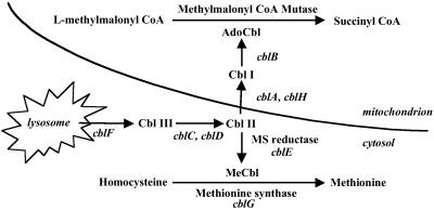
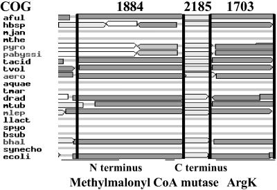
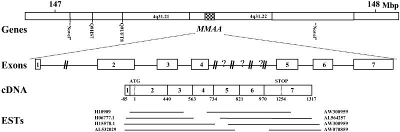
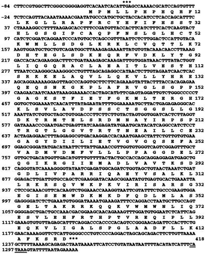
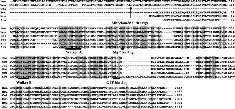
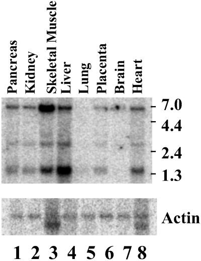
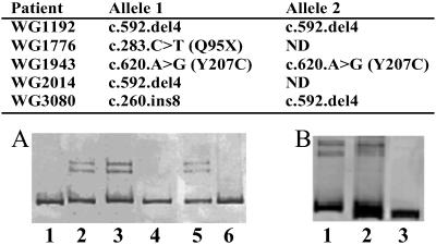
Vitamin B(12) (cobalamin) is an essential cofactor of two enzymes, methionine synthase and methylmalonyl-CoA mutase. The conversion of the vitamin to its coenzymes requires a series of biochemical modifications for which several genetic diseases are known, comprising eight complementation groups (cblA through cblH). The objective of this study was to clone the gene responsible for the cblA complementation group thought to represent a mitochondrial cobalamin reductase. Examination of bacterial operons containing genes in close proximity to the gene for methylmalonyl-CoA mutase and searching for orthologous sequences in the human genome yielded potential candidates. A candidate gene was evaluated for deleterious mutations in cblA patient cell lines, which revealed a 4-bp deletion in three cell lines, as well as an 8-bp insertion and point mutations causing a stop codon and an amino acid substitution. These data confirm that the identified gene, MMAA, corresponds to the cblA complementation group. It is located on chromosome 4q31.1-2 and encodes a predicted protein of 418 aa. A Northern blot revealed RNA species of 1.4, 2.6, and 5.5 kb predominating in liver and skeletal muscle. The deduced amino acid sequence reveals a domain structure, which belongs to the AAA ATPase superfamily that encompasses a wide variety of proteins including ATP-binding cassette transporter accessory proteins that bind ATP and GTP. We speculate that we have identified a component of a transporter or an accessory protein that is involved in the translocation of vitamin B(12) into mitochondria.


Figures

Fig 1.
Fig 1.
Intracellular processing of Cbl (modified from ref. 2). Steps are identified according to complementation group names. Their gene (uppercase italics) and predicted functions are: cblA (MMAA), this study; cblB, cob(I)alamin adenosyltransferase; cblC/D, possibly cob(III)alamin reductase; cblE (MTRR), methionine synthase reductase; cblF, lysosomal Cbl efflux; cblG (MTR), methionine synthase; and cblH, possibly a component of Cbl transport or cob(II)alamin reductase.
Fig 2.
Fig 2.
Cluster of orthologous genes examining the MCM N terminus (COG1884) and C terminus (COG2185) and the genes identified as comprising COG1703. Bacteria discussed in this study include Archaeoglobus fulgidus (aful) and Escherichia coli K12 (ecol); others are listed at www.ncbi.nlm.nih.gov/COG.
Fig 3.
Fig 3.
Structural organization of the human MMAA gene. The top schematic (“Genes”) illustrates the flanking genes and map positions surrounding MMAA according to contigview in Ensembl (www.ensembl.org). Note that NCBI maps MMAA to the 147.36–147.38 megabase pairs (Mbp) location of chromosome 4. Both Ensembl and NCBI localize MMAA to 4q31.21–22. The next line (“Exons”) illustrates the location of exons (numbered boxes) and introns (lines) along the MMAA gene located in Contig NT_016606.11. Our independently confirmed sequences from genomic DNA were deposited in GenBank under the accession nos. –. Introns are drawn to scale, except where “?” is used to denote a region of unknown sequence and length and “//” is used to denote a longer region than illustrated. Next the cDNA (“cDNA”) is illustrated, with splice and start/stop codons indicated, showing that it was constructed from the numerous ESTs (“ESTs”) belonging to Unigene clusters Hs.126261 (10 ESTs) and Hs.21017 (18 ESTs) and verified by RT-PCR sequencing. Only some ESTs are illustrated here.
Fig 4.
Fig 4.
Nucleotide and deduced amino acid sequence of human MMAA. The nucleotide and amino acid residues are numbered on the left and right margins, respectively. Two in-frame stop codons are indicated by *** immediately upstream and downstream of the coding sequence. A candidate polyA signal starting at 1,296 is underlined.
Fig 5.
Fig 5.
Alignment of amino acid sequences similar to that of human (Hum) MMAA, including mouse (Mou), Caenorhabditis elegans (Cel), E. coli (Eco), Mycobacterium tuberculosis (Mtu), and A. fulgidus (Afu). Sequence motifs defining the G3E family of proteins are indicated, including the GxxGxGK[S/T] Walker A, the Mg2+-binding aspartate residue, the DHbHbHbHbE Walker B motif with Hb denoting hydrophobic residues, and an [N/T]KxD GTP-binding motif, as described by Leipe et al. (27). The arrow denotes the predicted mitochondrial leader sequence cleavage site. Amino acids that are similar in all species are shaded with a gray background.
Fig 6.
Fig 6.
Northern blot analysis of MMAA mRNA. A Northern blot of poly(A)+ RNA from the indicated human tissues. The membranes were hybridized with a 32P-labeled 508-bp probe from MMAA mRNA (A) or a β-actin probe (B). Molecular size marker positions are indicated on the right.
Fig 7.
Fig 7.
Mutations reported here and heteroduplex tests. (A) Heteroduplex test for c.592.delACTG: lane 1, control cells; lane 2, WG2014 mixed with control cells in a 1:1 ratio; lane 3, WG2014 alone; lane 4, WG1192 alone; lane 5, WG1192 mixed with control; lane 6, WG1943 mixed with control cells in a 1:1 ratio. (B) Heteroduplex for c.259.delATAAACTT: lane 1, WG3080 alone; lane 2, WG3080 and control cells; lane 3, control cells alone.

References

    1. Raux E., Schubert, H. L. & Warren, M. J. (2000) Cell. Mol. Life Sci. 57 1880-1893. - PMC - PubMed
    1. Rosenblatt D. S. & Fenton, W. A. (2001) in The Metabolic and Molecular Bases of Inherited Disease, eds. Scriver, C. R., Beaudet, A. L., Valle, D. & Sly, W. S. (McGraw–Hill, New York), pp. 3897–3933.
    1. Leclerc D., Odievre, M., Wu, Q., Wilson, A., Huizenga, J. J., Rozen, R., Scherer, S. W. & Gravel, R. A. (1999) Gene 240 75-88. - PubMed
    1. Leclerc D., Campeau, E., Goyette, P., Adjalla, C. E., Christensen, B., Ross, M., Eydoux, P., Rosenblatt, D. S., Rozen, R. & Gravel, R. A. (1996) Hum. Mol. Genet. 5 1867-1874. - PubMed
    1. Li Y. N., Gulati, S., Baker, P. J., Brody, L. C., Banerjee, R. & Kruger, W. D. (1996) Hum. Mol. Genet. 5 1851-1858. - PubMed

Publication types

MeSH terms