. 2002 Nov 26;99(24):15393-7.
doi: 10.1073/pnas.232442599. Epub 2002 Nov 18.

Mitotic chromosomes are chromatin networks without a mechanically contiguous protein scaffold

Affiliations

Mitotic chromosomes are chromatin networks without a mechanically contiguous protein scaffold

Michael G Poirier et al. Proc Natl Acad Sci U S A. .

Abstract

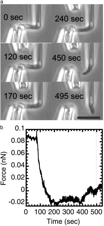
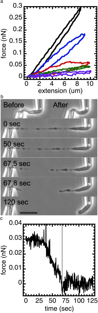
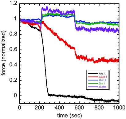
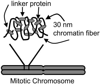
Isolated newt (Notophthalmus viridescens) chromosomes were studied by using micromechanical force measurement during nuclease digestion. Micrococcal nuclease and short-recognition-sequence blunt-cutting restriction enzymes first remove the native elastic response of, and then to go on to completely disintegrate, single metaphase newt chromosomes. These experiments rule out the possibility that the mitotic chromosome is based on a mechanically contiguous internal non-DNA (e.g., protein) "scaffold"; instead, the mechanical integrity of the metaphase chromosome is due to chromatin itself. Blunt-cutting restriction enzymes with longer recognition sequences only partially disassemble mitotic chromosomes and indicate that chromatin in metaphase chromosomes is constrained by isolated chromatin-crosslinking elements spaced by approximately 15 kb.


Figures

Fig 1.
Fig 1.
Mechanical response of a newt mitotic chromosome microdigested with 1 nM MN in 60% PBS with 1 mM CaCl2. The chromosome was put under 0.1 nN of force before microdigestion. MN causes a relaxation of the initially applied force before any apparent change in chromosome morphology in phase contrast microscopy. Longer exposures lead to dissolution of the chromosome into unobservably small fragments, showing that a large-scale protein scaffold does not exist within mitotic chromosomes. (a) Phase images of the chromosome being digested by MN. The time in each image corresponds to the time axis of b. (Bar = 10 μm.) A video is available at www.uic.edu/∼jmarko/published/enz. (b) Time series of the force supported by the chromosome during the nuclease digestion. The thin vertical line indicates the time at which the chromosome was severed. Digestion was initiated at t = 60 sec.
Fig 2.
Fig 2.
Partial digestion of a mitotic chromosome by MN shows it to behave as a chromatin network. A mitotic newt chromosome was digested with 10 nM MN for 90 sec without an applied tension. It was then subjected to four extension–retraction cycles in the absence of MN. The chromosome force constant was reduced after each extension–retraction cycle. After this, the mildly digested chromosome was extended to 40 μm. The chromosome does not elongate homogeneously; instead, there are blobs connected by thin fibers. To test whether the thin fibers contain DNA, we exposed the extended blob-thin fiber structure to 10 nM MN while monitoring the force. The force relaxes in response to the exposure and the blob-thin fiber structure is cut through, indicating that the thin fibers contain DNA and that it is required to support the applied tension. (a) Force–extension response of a chromosome before (black) and after a 90-sec chromosome digestion with 10 nM MN. After digestion (without an applied tension), successive extension–relaxation cycles (blue, red, green, and purple) progressively reduce the force response; there is no longer reversible elasticity. (b) Top images show the chromosome unextended before (Left) and after (Right) 90-sec MN microdigestion; there is no obvious change in morphology under zero force. Lower images labeled with a time show the final cutting of the chromosome extended to 40 μm, after the microdigestion and the extension–relaxation cycles of a. The t = 0 image shows the blob-link structure produced by microdigestion and stretching, whereas the t > 0 images show that the chromosome is completely cut by spraying with MN. (Bar = 10 μm.) (c) Time series of the digestion experiment of b. The chromosome was extended by ≈40 μm and supported a force of ≈30 pN. The microdigestion began at ≈40 sec. The force relaxes to zero as the chromosome is cut. The low levels of force in this experiment are insufficient to break a single DNA molecule. A video is available at www.uic.edu/∼jmarko/published/enz.
Fig 3.
Fig 3.
REs with increasing specificity show decreasing effects on chromosome elastic response. Force data are before, during, and after 350-sec exposures to various REs; force is normalized to units of initial applied force, which ranged between 0.2 and 0.8 nN in the five separate experiments shown. AluI AG↓CT (black) relaxes the force in ≈30 sec; Cac8I GCN↓NGC (red) only partially reduces the force. HincII GT(T/C)↓(A/G)AC (blue) and DraI TTT↓AAA (green) induce an increase in force during spraying, with a return to the original force when spraying stops (≈600 sec), similar to spraying with reaction buffer and no enzyme (violet). These results indicate that chromatin–chromatin crosslinks occur roughly every 15 kb (see text). A video of AluI digestion is available at www.uic.edu/∼jmarko/published/enz.
Fig 4.
Fig 4.
Proposed “network” model of higher mitotic chromosome structure. The black lines represent 30-nm chromatin fiber, and the gray ovals represent proteins connecting the chromatin fiber to form a network-type structure. This model has a structure where the proteins crosslink chromatin, which maintains higher chromosome structure. When this structure is exposed to MN, cuts in the DNA (chromatin) between the crosslinks will be induced. The chromosome will no longer support an applied tension, which is what is observed experimentally. In addition, we estimate the average number of base pairs between crosslinks to be ≈15 kb, based on the results from digesting the mitotic chromosome with various REs. Note that the crosslinks need not be homogeneously distributed through the chromatids.

Comment in

References

    1. Hirano T. (2000) Annu. Rev. Biochem. 69 115-144. - PubMed
    1. Dietzel S. & Belmont, A. S. (2001) Nat. Cell Biol. 3 767-770. - PubMed
    1. Paulson J. R. & Laemmli, U. K. (1977) Cell 12 817-828. - PubMed
    1. Marsden M. P. F. & Laemmli, U. K. (1979) Cell 17 849-858. - PubMed
    1. Laemmli U. K., Cheng, S. M., Adolph, K. W., Paulson, J. R., Brown, J. A. & Baumbach, W. R. (1978) Cold Spring Harbor Symp. Quant. Biol. 42 351-360. - PubMed

Publication types