. 2002 Nov 26;99(24):15422-7.
doi: 10.1073/pnas.222421399. Epub 2002 Nov 18.

Discovery of chemical inhibitors of the selective transfer of lipids mediated by the HDL receptor SR-BI

Affiliations

Discovery of chemical inhibitors of the selective transfer of lipids mediated by the HDL receptor SR-BI

Thomas J F Nieland et al. Proc Natl Acad Sci U S A. .

Abstract

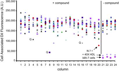
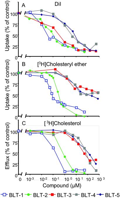
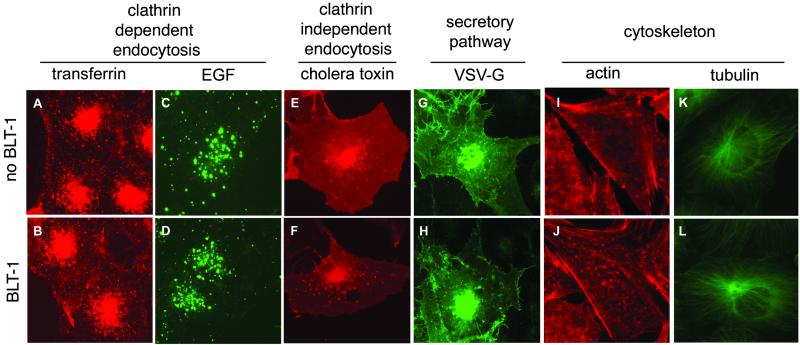
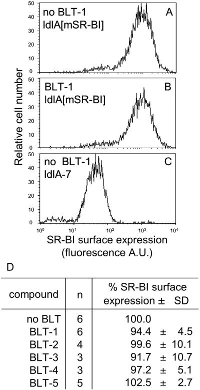
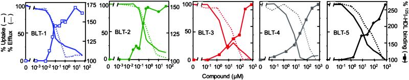
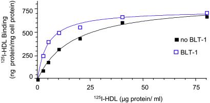
The high-density lipoprotein (HDL) receptor, scavenger receptor, class B, type I (SR-BI), mediates both the selective uptake of lipids, mainly cholesterol esters, from HDL to cells and the efflux of cholesterol from cells to lipoproteins. The mechanism underlying these lipid transfers is distinct from classic receptor-mediated endocytosis, but it remains poorly understood. To investigate SR-BI's mechanism of action and in vivo function, we developed a high-throughput screen to identify small molecule inhibitors of SR-BI-mediated lipid transfer in intact cells. We identified five compounds that in the low nanomolar to micromolar range block lipid transport (BLTs), both selective uptake and efflux. The effects of these compounds were highly specific to the SR-BI pathway, because they didn't interfere with receptor-mediated endocytosis or with other forms of intracellular vesicular traffic. Surprisingly, all five BLTs enhanced, rather than inhibited, HDL binding by increasing SR-BI's binding affinity for HDL (decreased dissociation rates). Thus, the BLTs provide strong evidence for a mechanistic coupling between HDL binding and lipid transport and may serve as a starting point for the development of pharmacologically useful modifiers of SR-BI activity and, thus, HDL metabolism.


Figures

Fig 1.
Fig 1.
High-throughput screen for inhibitors of SR-BI-mediated DiI uptake from DiI-HDL. Example of a fluorescent readout obtained from a single 384-well plate during the first round of the high-throughput screen. SR-BI-expressing ldlA[mSR-BI] cells were plated into 384-well plates, and the effect of compounds (≈10 μM) on the uptake of DiI from DiI-HDL (10 μg of protein per ml) was determined by using a high-speed fluorescence plate reader. Columns 1–20 show results (fluorescence in arbitrary units) from 16 independent wells per column (different colored symbols) from a single plate, representing a total of 320 compounds. Controls without compounds are wells containing ldlA[mSR-BI] cells in the absence or presence of a 40-fold excess of unlabeled HDL, or containing untransfected ldlA-7 cells (very low SR-BI expression). Wells containing an inhibitory compound named BLT-1 and wells with compounds that quenched DiI-HDL fluorescence (Q) are indicated.
Fig 2.
Fig 2.
Concentration dependence of the inhibition by BLTs of SR-BI-mediated lipid transfer between HDL and cells. ldlA[mSR-BI] cells were incubated with the indicated concentrations of BLTs, and their effects on DiI uptake from DiI-HDL (A), [3H]CE uptake from [3H]CE-HDL (B), and the efflux of [3H]cholesterol from cells to HDL (C) were determined. The 100% of control values were 50.6 ng of HDL protein equivalents per well (384-well plates) (A) and 3,908 ng of HDL protein equivalents per mg of cellular protein (B). In C, the data were normalized such that the maximum amount of [3H]cholesterol transferred from cells to HDL in the absence of compounds (55.7% of total) was set to 100%. The 0% value corresponds to the efflux of [3H]cholesterol transferred from ldlA[mSR-BI] cells to HDL without BLTs and in the presence of saturating inhibitory amounts of the specific anti-SR-BI blocking antibody KKB-1 (15% of total) (20). The efflux of [3H]cholesterol from ldlA-7 cells measured in the absence or presence of KKB-1 was 15% and 10% of total cellular [3H]cholesterol, respectively.
Fig 3.
Fig 3.
Effects of BLT-1 on intracellular membrane trafficking and cytoskeletal organization. Cells were incubated for 3 h in the absence (Upper) or presence (Lower) of 50 μM BLT-1, and epifluorescence light microscopy was used to monitor the following cellular activities: clathrin-dependent endocytosis of fluorescently labeled transferrin (A and B; HeLa cells) and epidermal growth factor (C and D; HeLa cells); clathrin-independent endocytosis of fluorescently labeled cholera toxin (E and F; BSC-1 cells); and transport of the temperature-sensitive fluorescent membrane protein VSV-Gts045-EGFP from the endoplasmic reticulum to the cell surface (G and H; BSC-1 cells). In addition, the intracellular distributions of the actin cytoskeleton (I and J; ldlA-[mSRBI] cells visualized with rhodamine-labeled phalloidin) and the tubulin network (K and L; BSC-1 cells visualized with fluorescently labeled antibodies specific to α-tubulin) were determined. BLT-1 and the other BLTs (data not shown) had no effects on any of these cellular properties or activities.
Fig 4.
Fig 4.
Cell surface expression of SR-BI. ldlA[mSR-BI] and ldlA-7 cells were treated for 3 h with or without BLTs at their corresponding IC95CE concentrations (1 μM for BLT-1 and BLT-2 and 50 μM for BLT-3–BLT-5) followed by determination of surface expression levels of SR-BI by flow cytometry. AC show histograms of the surface expression for ldlA[mSR-BI] cells without BLTs, ldlA[mSR-BI] cells with 1 μM BLT-1, and ldlA-7 cells without BLTs, respectively. D summarizes the results in ldlA[mSR-BI] cells for all five BLTs, with the value determined without compounds set to 100%. n, number of independent determinations.
Fig 5.
Fig 5.
Effects of BLTs on SR-BI-mediated cholesterol ether uptake from HDL, cellular cholesterol efflux to HDL, and HDL binding. The effects of the indicated concentrations of BLTs on SR-BI-mediated uptake of [3H]CE from [3H]CE-HDL (solid lines, no symbols), efflux of [3H]cholesterol from cells to HDL (dashed lines), or binding of 125I-HDL to cells (solid lines, symbols) were determined by using ldlA[mSR-BI] cells. To simplify comparisons, we define the lowest observed [3H]CE uptake and [3H]cholesterol efflux values (from Fig. 2) as 0% and the values in the absence of BLTs as 100%. The 100% control value for the 125I-HDL binding in the absence of BLTs was 403 ng of HDL protein per mg of cell protein.
Fig 6.
Fig 6.
Effects of BLT-1 on the concentration dependence of 125I-HDL binding to ldlA[mSR-BI] cells. The binding of 125I-HDL to ldlA[mSR-BI] cells was determined in duplicate at the indicated concentrations of HDL in the presence (blue) or absence (black) of 1 μM BLT-1 (IC95CE). Each value was corrected for binding of 125I-HDL in the presence of 40-fold excess of unlabeled HDL to ldlA [mSR-BI] cells in the presence of BLT-1.

References

    1. Acton S., Rigotti, A., Landschulz, K. T., Xu, S., Hobbs, H. H. & Krieger, M. (1996) Science 271 518-520. - PubMed
    1. Krieger M. (1999) Annu. Rev. Biochem. 68 523-558. - PubMed
    1. Trigatti B., Rayburn, H., Viñals, M., Braun, A., Miettinen, H., Penman, M., Hertz, M., Schrenzel, M., Amigo, L., Rigotti, A. & Krieger, M. (1999) Proc. Natl. Acad. Sci. USA 96 9322-9327. - PMC - PubMed
    1. Kozarsky K. F., Donahee, M. H., Glick, J. M., Krieger, M. & Rader, D. J. (2000) Arterioscler. Thromb. Vasc. Biol. 20 721-727. - PubMed
    1. Arai T., Wang, N., Bezouevski, M., Welch, C. & Tall, A. R. (1999) J. Biol. Chem. 274 2366-2371. - PubMed

Publication types

MeSH terms