

Carcinoma-like vascular density in atypic keratoacanthoma suggests malignant progression
- PMID: 12439721
- PMCID: PMC2408913
- DOI: 10.1038/sj.bjc.6600622
Carcinoma-like vascular density in atypic keratoacanthoma suggests malignant progression
Abstract
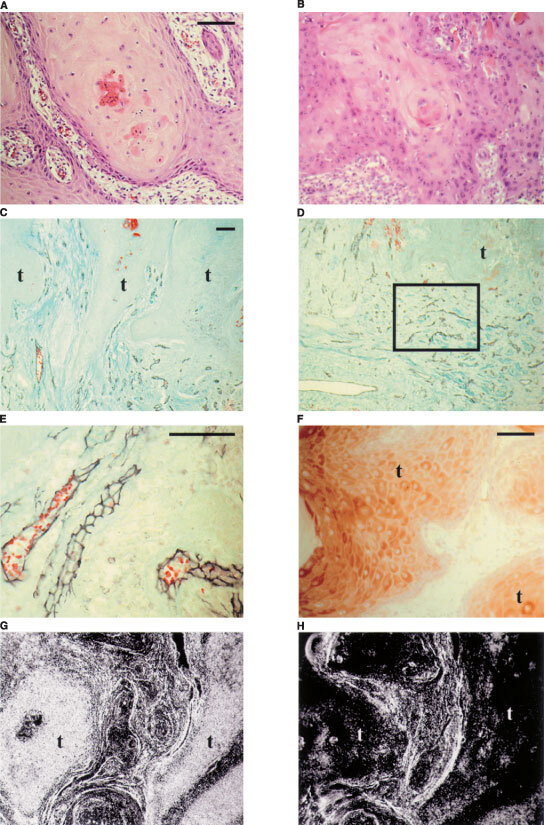
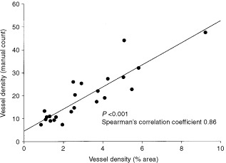
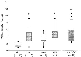
Differential diagnosis between keratoacanthomas and well differentiated squamous cell carcinomas based on clinical and histomorphological data is problematic. Recent findings of cellular atypia in a large proportion of keratoacanthomas indicated that these potentially 'self-healing' cutaneous neoplasms had the potential for malignant progression. Another malignancy-associated criterion is enhanced angiogenesis with increased microvessel density. To provide further diagnostic markers for keratoacanthomas we examined microvessel density on paraffin sections of 13 keratoacanthomas in comparison with 10 normal skin biopsies and 16 late-stage skin squamous cell carcinomas by counting and by computer-assisted image analysis of CD31-immunostained vessels. A significant increase of microvessel density in 'hot spots' was observed in keratoacanthomas as compared to normal skin. Furthermore, when keratoacanthomas were subdivided into tumours with and without malignancy-associated atypic areas, only those with atypia (n=6) were significantly better vascularised than normal skin and had a mean microvessel density in the range of late-stage squamous cell carcinomas. Both keratoacanthoma subtypes revealed comparable levels of inflammatory cell infiltration, tumour cell proliferation and vascular endothelial growth factor expression (mRNA and protein). Thus, in addition to malignancy-associated cellular atypia, increased microvessel density may serve as further diagnostic parameter to discriminate keratoacanthomas with a potential to progress to malignancy.
Figures
Similar articles
-
Differential expression of desmosomal glycoproteins in keratoacanthoma and squamous cell carcinoma of the skin: an immunohistochemical aid to diagnosis.Acta Derm Venereol. 1996 Sep;76(5):394-8. doi: 10.2340/0001555576394398. Acta Derm Venereol. 1996. PMID: 8891017
-
Transforming growth factor alpha expression helps to distinguish keratoacanthomas from squamous cell carcinomas.Arch Dermatol. 1991 Aug;127(8):1167-71. Arch Dermatol. 1991. PMID: 1863074
-
VCAM (CD-106) and ICAM (CD-54) adhesion molecules distinguish keratoacanthomas from cutaneous squamous cell carcinomas.Mod Pathol. 2003 Jan;16(1):8-13. doi: 10.1097/01.MP.0000043520.74056.CD. Mod Pathol. 2003. PMID: 12527707
-
Keratoacanthoma: hyperplasia, benign neoplasm, or a type of squamous cell carcinoma?Semin Diagn Pathol. 2009 Aug;26(3):150-63. doi: 10.1053/j.semdp.2009.09.003. Semin Diagn Pathol. 2009. PMID: 20043514 Review.
-
Differentiating keratoacanthoma from squamous cell carcinoma-In quest of the holy grail.J Cutan Pathol. 2020 Apr;47(4):418-420. doi: 10.1111/cup.13640. Epub 2020 Jan 14. J Cutan Pathol. 2020. PMID: 31893469 Review.
Cited by
-
Keratoacanthoma and Keratoacanthoma-Like Squamous Cell Carcinoma: Similar Morphology but Different Pathogenesis.Medicine (Baltimore). 2015 Jun;94(23):e934. doi: 10.1097/MD.0000000000000934. Medicine (Baltimore). 2015. PMID: 26061320 Free PMC article.
-
Patterns of LC3A Autophagy Protein Expression in Keratoacanthomas.Head Neck Pathol. 2020 Mar;14(1):150-155. doi: 10.1007/s12105-019-01033-1. Epub 2019 Apr 11. Head Neck Pathol. 2020. PMID: 30977096 Free PMC article.
References
-
- BarnetsonRSHallidayGM1997Regression in skin tumours: a common phenomenon Australas J Dermatol 38Suppl 1S63S65 - PubMed
-
- BillingsleyEMDavisNHelmKF1999Rapidly growing squamous cell carcinoma J Cutan Med Surg 3193197 - PubMed
-
- BreuningerHBlackBRassnerG1990Microstaging of squamous cell carcinomas Am J Clin Pathol 94624627 - PubMed
Publication types
MeSH terms
LinkOut - more resources
Full Text Sources
Medical
