

Structure determination of membrane proteins by NMR spectroscopy
- PMID: 12440700
- PMCID: PMC3454473
- DOI: 10.1139/o02-154
Structure determination of membrane proteins by NMR spectroscopy
Abstract
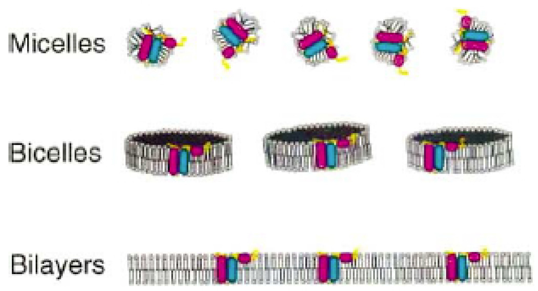
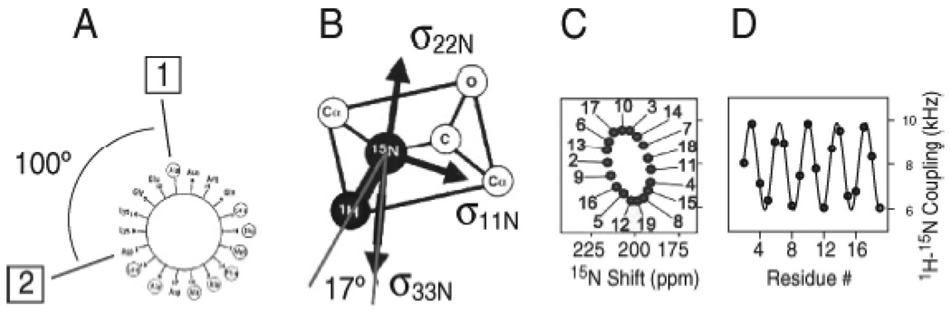
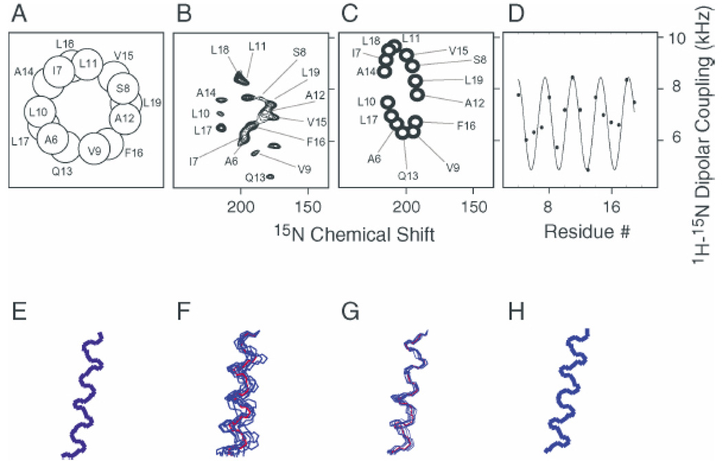
Current strategies for determining the structures of membrane proteins in lipid environments by NMR spectroscopy rely on the anisotropy of nuclear spin interactions, which are experimentally accessible through experiments performed on weakly and completely aligned samples. Importantly, the anisotropy of nuclear spin interactions results in a mapping of structure to the resonance frequencies and splittings observed in NMR spectra. Distinctive wheel-like patterns are observed in two-dimensional 1H-15N heteronuclear dipolar/15N chemical shift PISEMA (polarization inversion spin-exchange at the magic angle) spectra of helical membrane proteins in highly aligned lipid bilayer samples. One-dimensional dipolar waves are an extension of two-dimensional PISA (polarity index slant angle) wheels that map protein structures in NMR spectra of both weakly and completely aligned samples. Dipolar waves describe the periodic wave-like variations of the magnitudes of the heteronuclear dipolar couplings as a function of residue number in the absence of chemical shift effects. Since weakly aligned samples of proteins display these same effects, primarily as residual dipolar couplings, in solution NMR spectra, this represents a convergence of solid-state and solution NMR approaches to structure determination.
Figures
References
-
- Almeida FCL, Opella SJ. fd coat protein structure in membrane environments: structural dynamics of a loop connecting a hydrophobic trans-membrane helix and an amphiapathic helix in a membrane protein. J. Mol. Biol. 1997;270:481–495. - PubMed
-
- Bak M, Schultz R, Vosegaard T, Nielsen NC. Specification and visualization of anisotropic interaction tensors in polypeptides and numerical simulations in biological solid-state NMR. J. Magn. Reson. 2002;154:28–45. - PubMed
-
- Bax A, Kontaxis G, Tjandra N. Dipolar couplings in macromolecular structure determination. Methods Enzymol. 2001;330:127–172. - PubMed
-
- Cavanagh J, Fairbrother WJ, Palmer AG, Skelton NS. Protein NMR spectroscopy. New York: Academic Press; 1996.
-
- Chou JJ, Kaufman JD, Stahl SJ, Wingfield PT, Bax A. Micelle-induced curvature in a water-insoluble HIV-1 Env peptide revealed by NMR dipolar coupling measurement in stretched polyacrylamide gel. J. Am. Chem. Soc. 2002;124:2450–2451. - PubMed
Publication types
MeSH terms
Substances
Grants and funding
LinkOut - more resources
Full Text Sources
Other Literature Sources
