

The spinal antinociceptive effects of cholinergic drugs in rats: receptor subtype specificity in different nociceptive tests
- PMID: 12441008
- PMCID: PMC137595
- DOI: 10.1186/1471-2210-2-20
The spinal antinociceptive effects of cholinergic drugs in rats: receptor subtype specificity in different nociceptive tests
Abstract
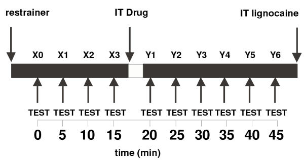
Background: Several studies have shown that muscarinic cholinergic agonists cause antinociception in humans and animals when given by both spinal and non-spinal parenteral routes. It is uncertain which subtype of muscarinic receptor is involved in spinally mediated antinociceptive effects caused by these drugs. The cholinergic receptor agonists McN-A-343 (M1 selective; 3.89 to 389 nmol) and carbachol (non-selective; 0.029 to 29 nmol) were used in a rat acute pain model to investigate the involvement of M1 and non-M1 subtypes in spinally mediated antinociception. The drugs were injected intrathecally and results from experiments in which drug actions were carefully confined to the spinal cord were used to construct agonist dose response curves.
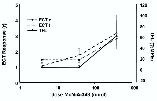
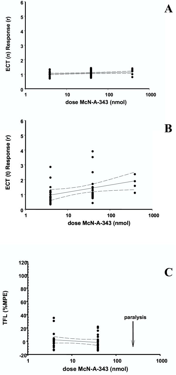
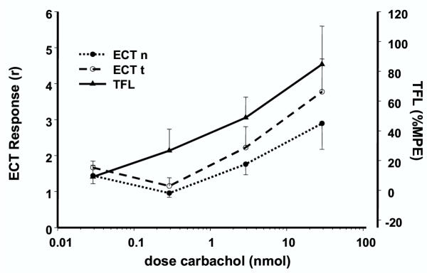
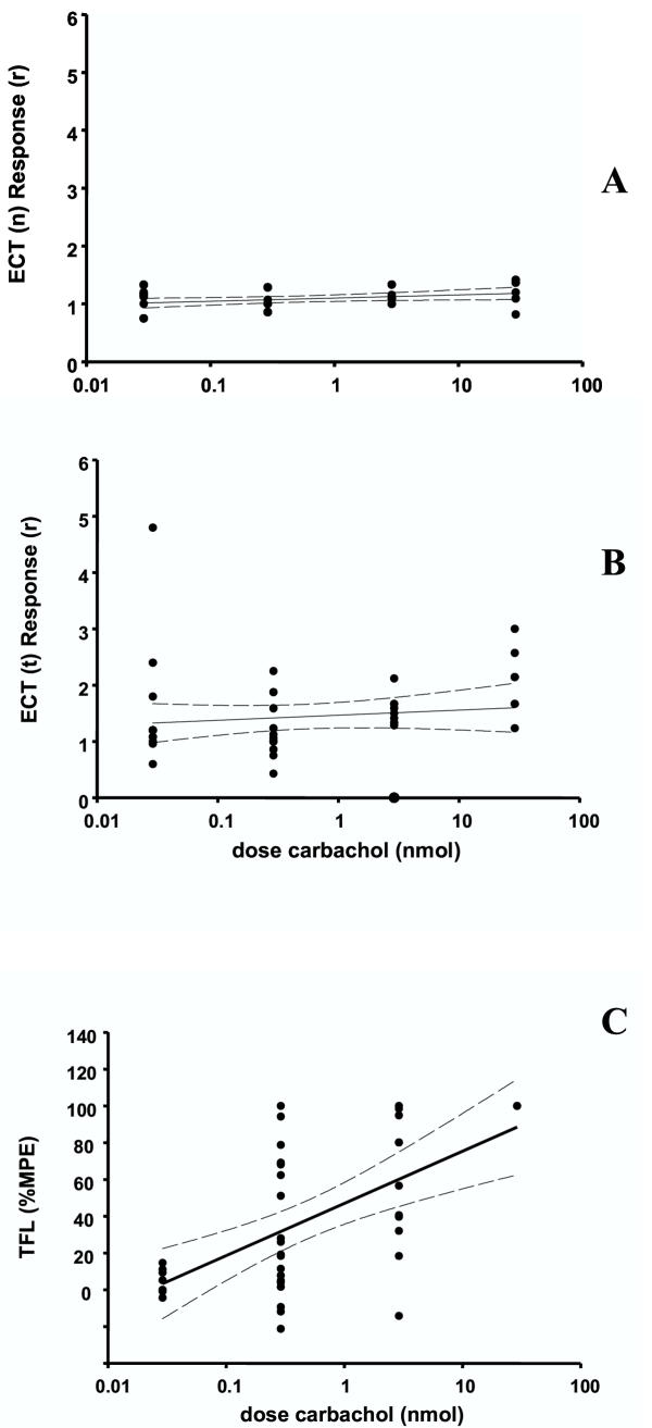
Results: McN-A-343 frequently diffused rostrally to the brain, away from the lumbosacral site of injection. Thus, in spite of its receptor subtype selectivity, McN-A-343 is a poor probe to use in attempting to identify receptor subtypes involved in spinal cord antinociceptive systems. However, in some experiments McN-A-343 caused spinally mediated antinociception assessed by the electrical current threshold test. Antinociception assessed by the tail flick latency test with intrathecal McN-A-343 was observed and found to involve supraspinal mechanisms. Carbachol caused spinally mediated antinociception assessed by both electrical current threshold and tail flick latency.
Conclusions: The results suggest that M1 receptors are involved in spinally mediated antinociception revealed by electrical current threshold; other cholinergic receptors (non-M1) are involved in thermal antinociception at the spinal cord. This contrasts with previous work on spinally mediated cholinergic antinociception. These differences are believed to be due to difficulties in restricting the action of these drugs to the spinal cord.
Figures
Similar articles
-
Activations of muscarinic M1 receptors in the anterior cingulate cortex contribute to the antinociceptive effect via GABAergic transmission.Mol Pain. 2017 Jan;13:1744806917692330. doi: 10.1177/1744806917692330. Mol Pain. 2017. PMID: 28326934 Free PMC article.
-
Antinociceptive role of 5-HT1A receptors in rat spinal cord.Br J Anaesth. 2002 May;88(5):679-84. doi: 10.1093/bja/88.5.679. Br J Anaesth. 2002. PMID: 12067006
-
The spinal muscarinic M(1) receptors and GABA(A) receptors contribute to the McN-A-343-induced antinociceptive effects during thermal stimulation of mice.J Pharmacol Sci. 2008 Dec;108(4):472-9. doi: 10.1254/jphs.08226fp. Epub 2008 Dec 5. J Pharmacol Sci. 2008. PMID: 19057125
-
Central antinociceptive effects of non-steroidal anti-inflammatory drugs and paracetamol. Experimental studies in the rat.Acta Anaesthesiol Scand Suppl. 1995;103:1-44. Acta Anaesthesiol Scand Suppl. 1995. PMID: 7725891 Review.
-
The pharmacology of McN-A-343.Pharmacol Ther. 2012 Aug;135(2):216-45. doi: 10.1016/j.pharmthera.2012.05.008. Epub 2012 May 27. Pharmacol Ther. 2012. PMID: 22643681 Review.
Cited by
-
Spinal cholinergic mechanism of the relieving effects of electroacupuncture on cold and warm allodynia in a rat model of neuropathic pain.J Physiol Sci. 2009 Jul;59(4):291-8. doi: 10.1007/s12576-009-0035-9. Epub 2009 Apr 3. J Physiol Sci. 2009. PMID: 19343482 Free PMC article.
-
Spinal microglial motility is independent of neuronal activity and plasticity in adult mice.Mol Pain. 2010 Apr 9;6:19. doi: 10.1186/1744-8069-6-19. Mol Pain. 2010. PMID: 20380706 Free PMC article.
-
Epidural administration of neostigmine-loaded nanofibers provides extended analgesia in rats.Daru. 2014 Nov 18;22(1):73. doi: 10.1186/s40199-014-0073-6. Daru. 2014. PMID: 25403313 Free PMC article.
References
-
- Chen G. The anti tremorine effect of some drugs as determined by Hoffner's method of testing analgesia in mice. J Pharmacol Exp Ther. 1958;124:73–76. - PubMed
-
- Herz A. Actions of arecoline on the central nervous system. Naunyn-Schmiedebergs Arch Exp Pathol Pharmacol. 1962;242:414–429. - PubMed
-
- Harris LS, Dewey WL, Howes JF, Kennedy JS, Pars H. Narcotic antagonist analgesics:interactions with cholinergic systems. J Pharmacol Exp Ther. 1969;169:17–22. - PubMed
MeSH terms
Substances
LinkOut - more resources
Full Text Sources
Medical
