. 2002 Dec 10;99(25):16297-302.
doi: 10.1073/pnas.252339399. Epub 2002 Nov 20.

Molecular basis of cell migration in the fish lateral line: role of the chemokine receptor CXCR4 and of its ligand, SDF1

Affiliations

Molecular basis of cell migration in the fish lateral line: role of the chemokine receptor CXCR4 and of its ligand, SDF1

Nicolas B David et al. Proc Natl Acad Sci U S A. .

Abstract

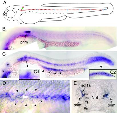
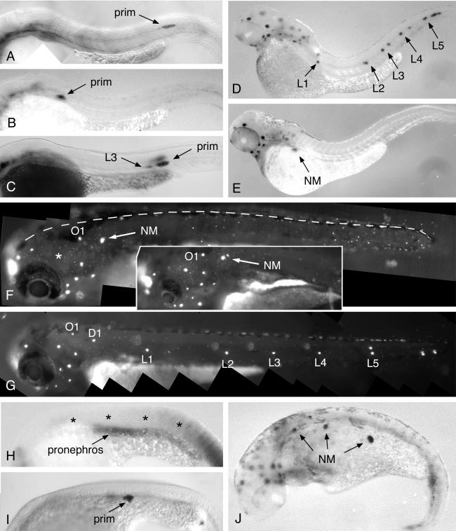
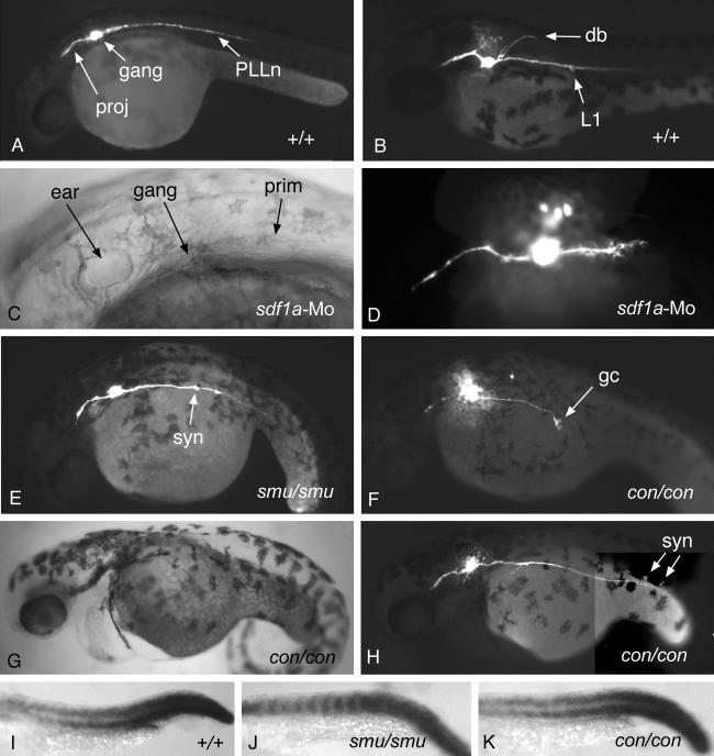
Cell migration plays an essential role in many morphogenetic processes, and its deregulation has many dramatic consequences. Yet how migration is controlled during normal development is still a largely unresolved question. We examined this process in the case of the posterior lateral line (PLL), a mechanosensory system present in fish and amphibians. In zebrafish, the embryonic PLL comprises seven to eight sense organs (neuromasts) aligned from head to tail along the flank of the animal and is formed by a primordium that originates from a cephalic placode. This primordium migrates along a stereotyped pathway toward the tip of the tail and deposits in its wake discrete groups of cells, each of which will become a neuromast. We show that a trail of SDF1-like chemokine is present along the pathway of the primordium and that a CXCR4-like chemokine receptor is expressed by the migrating cells. The inactivation of either the ligand or its receptor blocks migration, whereas in mutants in which the normal SDF1 trail is absent, the primordium path is redirected to the next, more ventral sdf1 expression domain. In all cases, the sensory axons remain associated to the primordium, indicating that the extension of the neurites to form the PLL nerve depends on the movement of the primordium. We conclude that both the formation and the innervation of this system depend on the SDF1-CXCR4 system, which has also been implicated in several migration events in humans, including metastasis formation and lymphocyte homing.


Figures

Fig 1.
Fig 1.
Migration of the PLL primordium. (A) In a 2-day-old embryo, the neuromasts of the PLL (blue dots) are deposited by a primordium that migrates along a stereotyped path (red dotted line). The first neuromast of the dorsal branch (green dot) has just formed; this line will later extend toward and along the dorsal midline (red arrowhead). (B) At 24 h postfertilization (hpf), the gene cxcr4b is predominantly expressed in the migrating primordium. (C) At the same time, the gene sdf1a is predominantly expressed in a stripe of cells that marks the pathway that the primordium will follow. Labeling is also observed at the level of the pronephros (arrowheads), rhombomeres 4 and 6 (asterisks), and in the forebrain. (C1) At 29 hpf, there is a marked expression of sdf1a at the level of somite 1, prefiguring the path of the dorsal branch. (C2) At 29 hpf, the posterior expression of sdf1a has narrowed down to the ventral portion of the terminal somites. (D) The PLL primordium (outlined by arrowheads) follows precisely the path marked by sdf1a-expressing cells in its posterior-wards migration (gray arrow). (E) In a section at the level of the migrating primordia (prim), the expression of sdf1a (blue) corresponds to a subset of myoseptal engrailed-expressing cells (En, gray nuclei). Not, notochord.
Fig 2.
Fig 2.
Morphants and mutants for sdf1a and cxcr4b. (A) The position of the PLL primordium ≈32 hpf in a normal embryo. The primordium has reached the level of the anus and is approximately halfway through its migration. In this and the following panels, the primordium is labeled by the cxcr4b probe. (B) In a sdf1a morphant (where sdf1a has been inactivated by morpholino-antisense injection), the primordium has barely moved at 32 hpf. (C) In a control embryo injected with a different morpholino, the primordium migrates normally. In this particular embryo, a group of cells is being deposited; note that cxcr4b is clearly down-regulated in these cells. (D) Pattern of neuromasts in a 2-day-old normal embryo. The neuromasts of the PLL are marked L1–L5; the two terminal neuromasts are not marked. (E) In a 2-day-old sdf1a morphant embryo, a single neuromast (NM) has formed at an abnormal, very anterior position. The head neuromasts show a normal pattern. In both D and E, neuromasts are stained with a specific probe (see Materials and Methods). (F) A 3-day-old cxcr4b morphant embryo labeled with 4-(4-diethylaminostyryl)-N-methylpyridinium iodide (4-Di-2-Asp), a fluorescent marker that specifically labels the hair cells of the neuromasts. The PLL is reduced to a single large neuromast behind the ear (NM); the occipital neuromast (O1) as well as the head neuromasts have formed normally. In this particular embryo, one supraorbital neuromast is missing (asterisk). The dorsal midline is marked by a dashed line. (Inset) Anterior one-half of another 3-day-old cxcr4b morphant embryo with a small eye; in this embryo, the PLL was reduced to two closely apposed, very anterior neuromasts (NM). In both cases, the “NM” neuromasts are located just posterior to the ear and anterior to the first somite, near the position occupied by the first neuromast of the dorsal line, D1, in normal embryos. (G) A 3-day-old control embryo that had been injected with a morpholino that recognizes the cxcr4b sequence just downstream of the start ATG. The PLL comprises five neuromasts aligned along the horizontal myoseptum, L1–L5, two terminal neuromasts at the tip of the tail, and the first neuromast of the dorsal line, D1. The occipital neuromast O1 has also been indicated to facilitate the comparison with the morphant embryos. (H) The smu mutant embryos lack a horizontal myoseptum and loose the lateral stripe of sdf1a expression (asterisks, compare to Fig. 1C). The expression of sdf1a is maintained at the level of the pronephros. (I) In a 32-hpf smu embryo, the PLL primordium (labeled with the cxcr4b probe) migrates along an ectopic ventral course that corresponds to the region where sdf1a is expressed. However, the movement is much slower than normal; compare to A. (J) In a 72-hpf smu embryo, three to four neuromasts are deposited and form an ectopic, ventral PLL. Same probe as in C and D.
Fig 3.
Fig 3.
Sensory neurons in morphants and mutants. (A) Application of the lipophilic neuronal tracer DiI in the PLL ganglion (gang), in fixed 32-hpf embryos. The central projection in the hindbrain (proj) and peripheral nerve extending to the primordium (PLLn) are labeled. (B) At 52 hpf, a synaptic specialization has developed under the L1 neuromast and the dorsal branch (db) has formed. (C) In a 32-hpf sdf1a morphant, the primordium (prim) has remained immobile, just posterior to the ganglion (gang). The otic vesicle (ear) has developed normally. (D) In the same embryo, the sensory neurons have extended a normal projection in the hindbrain, just beneath the ear, but their peripheral neurites have not extended beyond the primordium, where they branch profusely. (E) In a 32-hpf smu embryo, where the primordium travels along an ectopic ventral course, the sensory neurites follow this new pathway. A synapse with an ectopic neuromast (syn) is beginning to form in this particular embryo. (F) In chameleon (con) embryos, the horizontal myoseptum also fails to form and the corresponding stripe of sdf1a expression is missing. As in smu embryos, the sensory neurites accompany the primordium along its new course. gc, growth cones extending in the migrating primordium. (G) A 52-hpf con embryo. (H) The PLL neurons labeled in this embryo extend along the ectopic, ventral course of the primordium and establish synapses (syn) with two “terminal” neuromasts. (I) Expression of semaZ1a in a 24-hpf WT embryo. The gene is expressed in two broad domains dorsal and ventral to the horizontal myoseptum, as described (6). The expression has disappeared by 30 hpf (not shown). (J) In a 24-hpf smu embryo, the expression of semaZ1a does not show the normal gap at the level of the horizontal myoseptum. (K) In con embryos, however, the pattern of semaZ1a expression is not detectably modified.

References

    1. Dijkgraaf S. (1989) in The Mechanosensory Lateral Line: Neurobiology and Evolution, eds. Coombs, S., Görner, P. & Münz, H. (Springer, New York), pp. 7–14.
    1. Metcalfe W. K. (1989) J. Comp. Neurol. 238, 218-224. - PubMed
    1. Ledent V. (2002) Development (Cambridge, U.K.) 129, 597-604. - PubMed
    1. Sapède D., Gompel, N., Dambly-Chaudière, C. & Ghysen, A. (2002) Development (Cambridge, U.K.) 129, 605-615. - PubMed
    1. Gompel N., Cubedo, N., Thisse, C., Thisse, B., Dambly-Chaudière, C. & Ghysen, A. (2001) Mech. Dev. 105, 69-77. - PubMed

Publication types