

A developing picture of lymphopoiesis in bone marrow
- PMID: 12445263
- PMCID: PMC1850235
- DOI: 10.1034/j.1600-065x.2002.18904.x
A developing picture of lymphopoiesis in bone marrow
Abstract
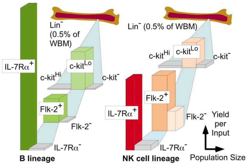
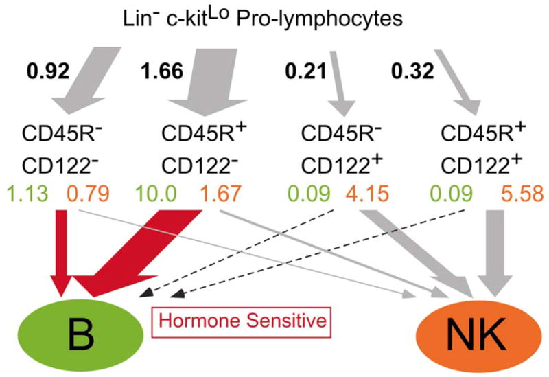
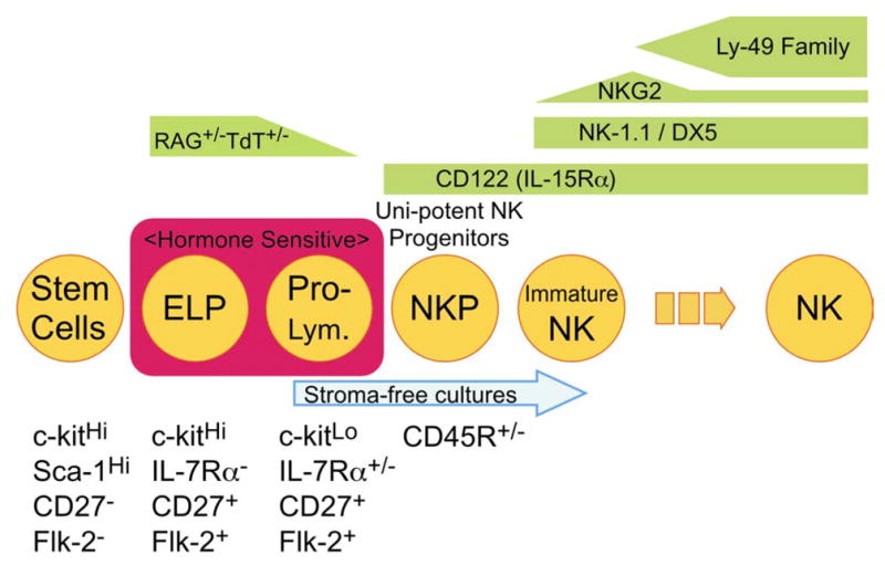
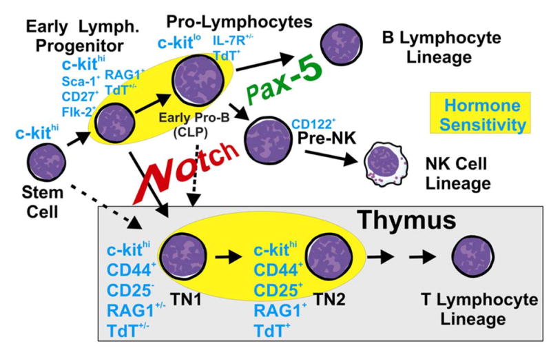
The earliest progenitors of lymphocytes are extremely rare and typically present among very complex populations of hematopoietic cells. Additionally, it is difficult to know how cells with any given set of characteristics are developmentally related to stem cells and maturing lymphoid precursors. However, it is now possible to divide bone marrow into progressively smaller fractions and exploit well-defined culture systems to determine which ones contain cells that can turn into lymphocytes. Analysis of steroid hormone sensitive cells and use of two-step cultures is providing additional information about the most likely differentiation pathways for B and natural killer cell lineage lymphocytes. A newly identified category of early lymphoid progenitors can now be sorted to high purity from RAG1/GFP knock in mice. Furthermore, the same experimental model makes it possible to image lymphoid progenitors in fetal and adult hematopoietic tissues.
Figures
Similar articles
-
Transcription from the RAG1 locus marks the earliest lymphocyte progenitors in bone marrow.Immunity. 2002 Aug;17(2):117-30. doi: 10.1016/s1074-7613(02)00366-7. Immunity. 2002. PMID: 12196284
-
Asynchronous RAG-1 expression during B lymphopoiesis.J Immunol. 2009 Dec 15;183(12):7768-77. doi: 10.4049/jimmunol.0902333. J Immunol. 2009. PMID: 20007571 Free PMC article.
-
Suppression of B lymphopoiesis at a lymphoid progenitor stage in adult rabbits.Int Immunol. 2007 Jun;19(6):801-11. doi: 10.1093/intimm/dxm048. Epub 2007 May 13. Int Immunol. 2007. PMID: 17502309
-
[Lymphopoiesis supported by osteolineage cells].Clin Calcium. 2016 May;26(5):707-12. Clin Calcium. 2016. PMID: 27117616 Review. Japanese.
-
Trafficking from the bone marrow to the thymus: a prerequisite for thymopoiesis.Immunol Rev. 2006 Feb;209:47-57. doi: 10.1111/j.0105-2896.2006.00350.x. Immunol Rev. 2006. PMID: 16448533 Review.
Cited by
-
Ageing, autoimmunity and arthritis: senescence of the B cell compartment - implications for humoral immunity.Arthritis Res Ther. 2004;6(4):131-9. doi: 10.1186/ar1180. Epub 2004 May 10. Arthritis Res Ther. 2004. PMID: 15225355 Free PMC article. Review.
-
Multiparameter Flow Cytometry to Detect Hematogones and to Assess B-lymphocyte clonality in Bone Marrow Samples from Patients with Non-Hodgkin Lymphomas.Hematol Rep. 2014 Jun 26;6(2):5381. doi: 10.4081/hr.2014.5381. eCollection 2014 Apr 22. Hematol Rep. 2014. PMID: 25013717 Free PMC article.
-
Wnt expression and canonical Wnt signaling in human bone marrow B lymphopoiesis.BMC Immunol. 2006 Jun 29;7:13. doi: 10.1186/1471-2172-7-13. BMC Immunol. 2006. PMID: 16808837 Free PMC article.
-
Aging, B lymphopoiesis, and patterns of leukemogenesis.Exp Gerontol. 2007 May;42(5):391-5. doi: 10.1016/j.exger.2006.11.010. Epub 2006 Dec 20. Exp Gerontol. 2007. PMID: 17184948 Free PMC article. Review.
-
Clinical Predictors for Neutrophil-to-Lymphocyte Ratio Changes in Patients with Chronic Hepatitis B Receiving Peginterferon Treatment.In Vivo. 2017 Jul-Aug;31(4):723-729. doi: 10.21873/invivo.11121. In Vivo. 2017. PMID: 28652447 Free PMC article.
References
-
- Kincade PW, Phillips RA. B lymphocyte development. Fed Proc. 1985;44:2874–2881. - PubMed
-
- Lemischka IR, Raulet DH, Mulligan RC. Developmental potential and dynamic behavior of hemopoietic stem cells. Cell. 1986;45:917–927. - PubMed
-
- Kondo M, Weissman IL, Akashi K. Identification of clonogenic common lymphoid progenitors in mouse bone marrow. Cell. 1997;91:661–672. - PubMed
-
- Roitt IM, Brostoff J, Male D. Immunology. St. Louis; C.V. Mosby: 1985.
Publication types
MeSH terms
Substances
Grants and funding
LinkOut - more resources
Full Text Sources
