. 2002 Dec;3(12):1222-7.
doi: 10.1093/embo-reports/kvf239. Epub 2002 Nov 21.

Role of ubiquitination in retro-translocation of cholera toxin and escape of cytosolic degradation

Affiliations

Role of ubiquitination in retro-translocation of cholera toxin and escape of cytosolic degradation

Chiara Rodighiero et al. EMBO Rep. 2002 Dec.

Abstract

Cholera toxin travels from the cell surface of affected mammalian cells to the endoplasmic reticulum (ER), where the A1 chain is released and retro-translocated across the ER membrane into the cytosol. We have tested whether, as in other cases, retro-translocation requires poly-ubiquitination. We show that an A1 chain mutant that lacks lysines and has a blocked N-terminus, and therefore cannot be ubiquitinated, remains active in vivo. The A1 chain is not degraded in the cytosol, as demonstrated by the fact that proteasome inhibitors do not stimulate its activity. When additional lysines are introduced into the A1 chain, moderate degradation by the proteasome is observed. The unfolded A1 chain rapidly refolds in vitro. These results show that poly-ubiquitination is not required for retro-translocation of all proteins across the ER membrane and indicate that the reason why the toxin escapes degradation in the cytosol may be both its paucity of lysines and its rapid refolding.


Figures
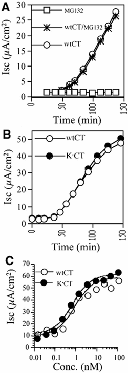
Figure 1
Figure 1
CT activity in vivo does not depend on poly-ubiquitination or proteasome function. (A) Time-course of CT-induced Cl secretion (Isc) in T84 cells treated with MG132/DMSO, DMSO alone or MG132 but not CT. (B) Time-course of Isc induced by 0.5 nM CT and the lysine-less KCT variant. (C) Dose response for peak Isc induced by CT or KCT.
Figure 2
Figure 2
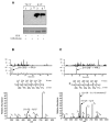
Complete blockade of the N-terminus of the CTA1 chain. (A) Upper: non-reducing SDS–PAGE and avidin-HRP blot of biotin-conjugated KCT blocked by carbamylation (asterisk, lanes 1 and 2) or unblocked (lanes 3 and 4). Biotinylation was performed in 0.4% SDS (lanes 1 and 3) or in aqueous buffer (lanes 2 and 4). Lower: Coomassie staining of an equivalent gel shows comparable sample loading. (B) The N-terminal peptide of the unblocked KCT was identified by mass spectroscopy analysis as a free amine. The doubly charged peptide ion (m/z = 476.6) was eluted early in the gradient, and the resulting tandem mass can be matched to the peptide sequence shown without modification. (C) The N-terminus in *KCT is blocked by carbamylation. The m/z = 476.6 peak in (B) is missing after carbamylation, and a new peak corresponding to the modified doubly charged peptide ion (m/z = 498.2) is detected with an increased retention time. The resulting tandem mass spectrum of this ion shows an N-terminus modified by the mass of a carbamylation (43 Da).
Figure 3
Figure 3
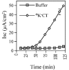
Poly-ubiquitination of CTA1 chain is not required for its retro-translocation. Time-course of Isc in T84 cells induced by 100 nM N-terminally blocked KCT (*KCT) or buffer alone.
Figure 4
Figure 4
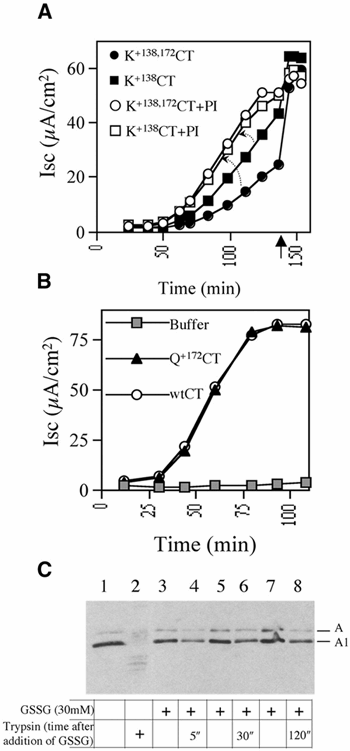
The CTA1 chain evades proteasome-dependent degradation by two mechanisms. (A) Isc induced by the lysine-rich CTA1 variants K+138CT or K+138,172CT in T84 monolayers treated with the proteasome inhibitor (PI) MG132/DMSO or DMSO alone. Forskolin (added at arrow) induces maximum Isc in all monolayers. (B) Time-course of Isc induced by 100 nM CT mutant with a glutamine substitution at position 172 in the CTA1 chain (Q+172CT), 100 nM CT or buffer alone. (C) Purified PDI was incubated in 1 mM GSH with isolated CTA subunit. Where indicated, 30 mM GSSG was added to release the A1 chain from PDI. At the indicated times, trypsin was added (lanes 2, 4, 6 and 8) or not added (lanes 1, 3, 5 and 7) to assay for the kinetics of A1 chain refolding. Lanes 1 and 2 show negative and positive control, respectively, with trypsin added 5 min after GSSG. All samples were analyzed by non-reducing SDS–PAGE and immunoblotting. The A subunit (A1 and A2 chains) and the reduced A1 chain are shown.

Similar articles

Cited by

References

    1. Biederer T., Volkwein C. and Sommer T. (1996) Degradation of subunits of the Sec61p complex, an integral component of the ER membrane, by the ubiquitin–proteasome pathway. EMBO J., 15, 2069–2076. - PMC - PubMed
    1. Breitschopf K., Bengal E., Ziv T., Admon A. and Ciechanover A. (1998) A novel site for ubiquitination: the N-terminal residue, and not internal lysines of MyoD, is essential for conjugation and degradation of the protein. EMBO J., 17, 5964–5973. - PMC - PubMed
    1. Deeks E.D., Cook J.P., Day P.J., Smith D.C., Roberts L.M. and Lord J.M. (2002) The low lysine content of ricin A chain reduces the risk of proteolytic degradation after translocation from the endoplasmic reticulum to the cytosol. Biochemistry, 41, 3405–3413. - PubMed
    1. Hazes B. and Read R.J. (1997) Accumulating evidence suggests that several AB-toxins subvert the endoplasmic reticulum-associated protein degradation pathway to enter target cells. Biochemistry, 36, 11051–11054. - PubMed
    1. Hershko A. and Ciechanover A. (1998) The ubiquitin system. Annu. Rev. Biochem., 67, 425–479. - PubMed

Publication types