. 2002 Nov 25;159(4):563-9.
doi: 10.1083/jcb.200208001.

Missense mutation in the tubulin-specific chaperone E (Tbce) gene in the mouse mutant progressive motor neuronopathy, a model of human motoneuron disease

Affiliations

Missense mutation in the tubulin-specific chaperone E (Tbce) gene in the mouse mutant progressive motor neuronopathy, a model of human motoneuron disease

Heike Bommel et al. J Cell Biol. .

Abstract

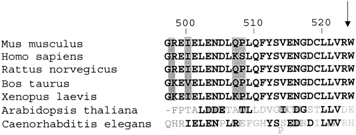
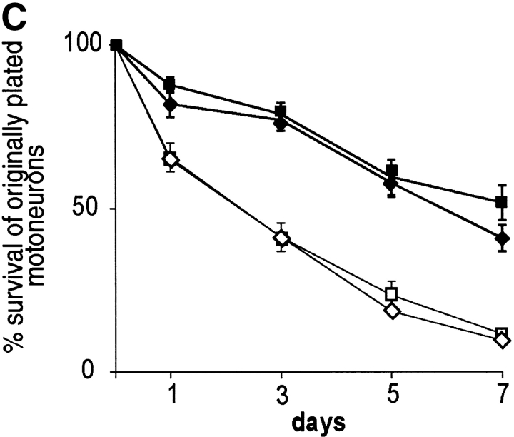
Progressive motor neuronopathy (pmn) mutant mice have been widely used as a model for human motoneuron disease. Mice that are homozygous for the pmn gene defect appear healthy at birth but develop progressive motoneuron disease, resulting in severe skeletal muscle weakness and respiratory failure by postnatal week 3. The disease starts at the motor endplates, and then leads to axonal loss and finally to apoptosis of the corresponding cell bodies. We localized the genetic defect in pmn mice to a missense mutation in the tubulin-specific chaperone E (Tbce) gene on mouse chromosome 13. The human orthologue maps to chromosome 1q42.3. The Tbce gene encodes a protein (cofactor E) that is essential for the formation of primary alpha-tubulin and beta-tubulin heterodimeric complexes. Isolated motoneurons from pmn mutant mice exhibit shorter axons and axonal swelling with irregularly structured beta-tubulin and tau immunoreactivity. Thus, the pmn gene mutation provides the first genetic evidence that alterations in tubulin assembly lead to retrograde degeneration of motor axons, ultimately resulting in motoneuron cell death.


Figures

Figure 1.
Figure 1.
Characterization of the gene defect in pmn mice. (A) Genetic map of the pmn candidate region. (B) Transcript map of the pmn region including the synteny region of the human genome. Transcriptional units not present in public databases were numbered according to their succession in this region. Pmn2 is located in noncoding regions of Nid. (C) Primary genomic DNA sequence of Tbce in pmn, heterozygous, and NMRI wt mice. Sequences of the sense strand are shown. The arrow indicates the T→G base mutation. The mutation leads to an exchange of the amino acid Trp to Gly. (D) Segregation of the homozygous Tbce G/Tbce G mutation with the pmn disease phenotype. One representative pedigree is shown. Homozygous Tbce G/Tbce G mutation was only found in pmn mice (black symbols). Healthy littermates were either heterozygous for the Tbce G mutation (Tbce W/Tbce G; gray symbols) or homozygous wt (Tbce W/Tbce W; white symbols). The animals marked with a black dot showed symptoms of motoneuron disease but died before genotyping was initiated.
Figure 2.
Figure 2.
Multiple amino acid sequence alignment of the COOH-terminal region of CofE from different vertebrate and nonvertebrate species. The nucleotide sequences of CofE were taken from public databases, translated to the corresponding amino acid sequence, and then aligned using ExPasy (www2.ebi.ac.uk/clustal). The amino acid numbers correspond to murine CofE. Conserved substitutions and semiconserved substitutions are indicated in gray; the remaining black amino acids are identical. Amino acids without similarity are shown in light gray. The arrow indicates the amino acid mutated in pmn mice.
Figure 3.
Figure 3.
Axonal growth and survival in isolated wt and pmn mutant motoneurons. (A and B) After 7 d in culture with 1 ng/ml BDNF, the length of the longest axon and the total length of the axon including all its branches from pmn mutant motoneurons (Tbce G/Tbce G; open bar) are significantly (P < 0.0001) shorter than from wt motoneurons (Tbce W/Tbce W; closed bar). (C) Survival of motoneurons isolated from wt (Tbce W/Tbce W; ♦) and pmn mice (Tbce G/Tbce G; ▪) in the presence (closed symbols) and absence (open symbol) of 1 ng/ml BDNF.
Figure 3.
Figure 3.
Axonal growth and survival in isolated wt and pmn mutant motoneurons. (A and B) After 7 d in culture with 1 ng/ml BDNF, the length of the longest axon and the total length of the axon including all its branches from pmn mutant motoneurons (Tbce G/Tbce G; open bar) are significantly (P < 0.0001) shorter than from wt motoneurons (Tbce W/Tbce W; closed bar). (C) Survival of motoneurons isolated from wt (Tbce W/Tbce W; ♦) and pmn mice (Tbce G/Tbce G; ▪) in the presence (closed symbols) and absence (open symbol) of 1 ng/ml BDNF.
Figure 4.
Figure 4.
Morphology of spinal motoneurons isolated from 13.5-d-old wt (TbceW/TbceW) and pmn (TbceG/TbceG) mutant embryos. Motoneurons were cultured for 7 d with 1 ng/ml BDNF, fixed, and stained against β III tubulin (red) and tau (green). (A and B) After 7 d in culture with 1 ng/ml BDNF, many Tbce G/Tbce G motoneurons exhibited axonal swellings (B, arrowheads), as defined by a locally restricted, irregular shaped, more than threefold increase in axon diameter. (A) wt (Tbce W/Tbce W) embryos. Bars, 50 μM. (C) Blinded analysis of motoneurons from Tbce W/Tbce W (n = 53) and Tbce G/Tbce G (n = 55) mutant embryos revealed a >20-fold increase in the number of motoneurons with this specific alteration.
Figure 5.
Figure 5.
Morphology of spinal motoneurons isolated from 13.5-d-old wt (TbceW/TbceW) and pmn mutant (TbceG/TbceG) embryos. Motoneurons were cultured for 7 d with 1 ng/ml BDNF, fixed, and stained against β III tubulin (red) and tau (green). (A and B) Motoneurons derived from pmn mutant embryos (B) exhibit shorter axons compared with axons from control cells (A). In pmn mutant motoneurons axonal varicosities are detectable (B, arrowhead), which were rarely observed in wt motoneurons. (C–H) Whereas β III tubulin and tau immunoreactivity is regularly organized and evenly distributed in axonal branch points in motoneurons derived from wt embryos (C), immunoreactivity for β III tubulin and tau in pmn mutants (D–H) is irregularly distributed in aggregate-like structures, and both proteins do not colocalize. (I–L) In contrast, growth cones that are mostly devoid of microtubular structures are morphologically indistinguishable in wt (I and J) and pmn mutant motoneurons (K and L). Bars: (A and B) 25 μm; (C, E, F, H) 5 μm; (D and G) 25 μm; (J and L) 5 μm; (I and K) 25 μm.

References

    1. Altschul, S.F., T.L. Madden, A.A. Schaffer, J. Zhang, Z. Zhang, W. Miller, and D.J. Lipman. 1997. Gapped BLAST and PSI-BLAST: a new generation of protein database search programs. Nucleic Acids Res. 25:3389–3402. - PMC - PubMed
    1. Bhamidipati, A., S.A. Lewis, and N.J. Cowan. 2000. ADP ribosylation factor-like protein 2 (Arl2) regulates the interaction of tubulin-folding cofactor D with native tubulin. J. Cell Biol. 149:1087–1096. - PMC - PubMed
    1. Cote, F., J.F. Collard, and J.P. Julien. 1993. Progressive neuronopathy in transgenic mice expressing the human neurofilament heavy gene: a mouse model of amyotrophic lateral sclerosis. Cell. 73:35–46. - PubMed
    1. Ferreira, A., and A. Caceres. 1992. Expression of the class III β-tubulin isotype in developing neurons in culture. J. Neurosci. Res. 32:516–529. - PubMed
    1. Garcia, M.L., and D.W. Cleveland. 2001. Going new places using an old MAP: tau, microtubules and human neurodegenerative disease. Curr. Opin. Cell Biol. 13:41–48. - PubMed

MeSH terms

Associated data