. 2002 Nov 25;159(4):601-11.
doi: 10.1083/jcb.200205084.

Iqg1p links spatial and secretion landmarks to polarity and cytokinesis

Affiliations

Iqg1p links spatial and secretion landmarks to polarity and cytokinesis

Mahasin A Osman et al. J Cell Biol. .

Abstract

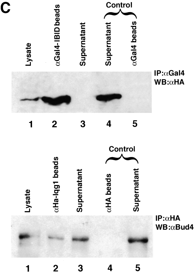
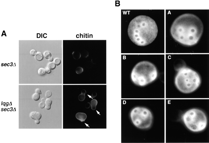
Cytokinesis requires the polarization of the actin cytoskeleton, the secretion machinery, and the correct positioning of the division axis. Budding yeast cells commit to their cytokinesis plane by choosing a bud site and polarizing their growth. Iqg1p (Cyk1p) was previously implicated in cytokinesis (Epp and Chant, 1997; Lippincott and Li, 1998; Osman and Cerione, 1998), as well as in the establishment of polarity and protein trafficking (Osman and Cerione, 1998). To better understand how Iqg1p influences these processes, we performed a two-hybrid screen and identified the spatial landmark Bud4p as a binding partner. Iqg1p can be coimmunoprecipitated with Bud4p, and Bud4p requires Iqg1p for its proper localization. Iqg1p also appears to specify axial bud-site selection and mediates the proper localization of the septin, Cdc12p, as well as binds and helps localize the secretion landmark, Sec3p. The double mutants iqg1Deltasec3Delta and bud4Deltasec3Delta display defects in polarity, budding pattern and cytokinesis, and electron microscopic studies reveal that these cells have aberrant septal deposition. Taken together, these findings suggest that Iqg1p recruits landmark proteins to form a targeting patch that coordinates axial budding with cytokinesis.


Figures

Figure 1.
Figure 1.
Iqg1p binds Bud4p. (A) Schematic representation of Bud4p. IBID, represents the Iqg1p-Bud4 Interacting Domain, amino acids 769–880. The RGD motif (a specific cell binding tri-peptide Arg-Gly-Asp) at position 819 is similar to that found in mammalian extracellular matrix proteins. GTP, stands for the GTP-binding and hydrolysis domain, amino acids 1175–1403 (Sanders and Herskowitz, 1996). PH, is a Pleckstrin Homology motif at amino acids 1304–1407. (B) Domains of IQG1 cloned into the two-hybrid plasmid pGBD-C2. (a) Schematic representation of Iqg1p domains. (b) The NH2 terminus (NG). (c) NH2 terminus (CG) lacking the Calponin Homology domain (CHD). (d) The COOH terminus of Iqg1p that includes the RasGAP-like domain. (C) Iqg1p coimmunoprecipitates (coIP) with Bud4p. (Top) MO3 cells were cotransformed with HA-IQG1 and GAL4IBID (the fragment containing the Iqg1p-binding domain) or, for control, with the Gal4-binding domain plasmid (empty vector) and the HA-IQG1 plasmid. Cells were grown to saturation in cm-leu-trp. The total cell lysate was used for coIP with the α-Gal4 binding domain antibody. Western blot analysis was performed using an α-HA antibody to detect HA-tagged Iqg1p. (Bottom) MO3 cells were transformed with HA-IQG1 plasmid or with empty vector as a control. Total cell lysate was used for coIP with α-HA antibodies and Western blot analysis was performed using affinity-purified α-Bud4p antibodies.
Figure 1.
Figure 1.
Iqg1p binds Bud4p. (A) Schematic representation of Bud4p. IBID, represents the Iqg1p-Bud4 Interacting Domain, amino acids 769–880. The RGD motif (a specific cell binding tri-peptide Arg-Gly-Asp) at position 819 is similar to that found in mammalian extracellular matrix proteins. GTP, stands for the GTP-binding and hydrolysis domain, amino acids 1175–1403 (Sanders and Herskowitz, 1996). PH, is a Pleckstrin Homology motif at amino acids 1304–1407. (B) Domains of IQG1 cloned into the two-hybrid plasmid pGBD-C2. (a) Schematic representation of Iqg1p domains. (b) The NH2 terminus (NG). (c) NH2 terminus (CG) lacking the Calponin Homology domain (CHD). (d) The COOH terminus of Iqg1p that includes the RasGAP-like domain. (C) Iqg1p coimmunoprecipitates (coIP) with Bud4p. (Top) MO3 cells were cotransformed with HA-IQG1 and GAL4IBID (the fragment containing the Iqg1p-binding domain) or, for control, with the Gal4-binding domain plasmid (empty vector) and the HA-IQG1 plasmid. Cells were grown to saturation in cm-leu-trp. The total cell lysate was used for coIP with the α-Gal4 binding domain antibody. Western blot analysis was performed using an α-HA antibody to detect HA-tagged Iqg1p. (Bottom) MO3 cells were transformed with HA-IQG1 plasmid or with empty vector as a control. Total cell lysate was used for coIP with α-HA antibodies and Western blot analysis was performed using affinity-purified α-Bud4p antibodies.
Figure 2.
Figure 2.
Iqg1p specifies axial budding and localizes Bud4p. (A) Iqg1p selects the axial bud-site: wild-type, bud4Δ, and iqg1Δ cells were grown in YEPD at 26°C, incubated with Calcofluor as described in Materials and methods, and photographed under identical conditions. (A–H) MO3 (iqg1Δ) cells. (B) Subcellular localization of Bud4p in wild-type and iqg1Δ cells: The subcellular localization of Bud4p was examined in wild-type IQG1 cells (MO5) and in iqg1Δ cells (MO3) as indicated in the panels. Cultures were grown at 23°C overnight, shifted to 30°C for 2 h, fixed with 3.7% formaldehyde and processed for indirect immunofluorescence using an affinity purified anti-Bud4p antibody as described in the Materials and methods.
Figure 3.
Figure 3.
Interactions between Iqg1p, Bud4p, and Cdc12p. (A) Examination of the interactions between Cdc12p and either Iqg1p or Bud4p by coimmunoprecipitation. MO3 cells were transformed with CDC12-GFP on a low-copy vector and HA-IQG1 plasmids. Cells were grown to saturation in media lacking uracil and leucine or with GFP empty vector along with the HA-IQG1 plasmid. The α-GFP antibodies were used to coIP Cdc12p-GFP from total cell lysates. Western blot analysis was performed using α-HA antibodies (top) to detect HA-tagged Iqg1p or affinity-purified α-Bud4p antibodies to detect the endogenous Bud4p (lower panel). The α-Intersectin antibody was used as an additional control. (B) Localization of Cdc12p in wild-type and iqg1Δ cells. The subcellular location of a Cdc12p-GFP fusion protein was examined in wild-type IQG1 cells (MO1) and in iqg1Δ cells (MO4) as indicated above the panels. (Top) Cells that carried the CDC12-GFP gene on the low-copy vector YCplac111. (Bottom) Cells that carried the CDC12-GFP gene on the high-copy vector YEplac181. Cultures were grown overnight at 23°C, shifted to 30°C for 6 h, and then photographed. All photography and printing steps were performed under identical conditions to preserve the different intensities of Cdc12p-GFP fluorescence.
Figure 4.
Figure 4.
Iqg1p binds and helps localize Sec3p. (A) Iqg1p binds Sec3p. MO3 cells were cotransformed with HA-IQG1 and SEC3-GFP plasmids, or with HA-IQG1 and GFP empty vector, and grown to saturation. The total cell lysate was used to immunoprecipitate Sec3p-GFP using α-GFP antibodies along with other control antibodies (α-Intersectin and α-HA). The proteins were fractionated on a 7% SDS-PAGE and the immunoblot was stained with α-HA antibodies to detect HA-Iqg1p. (B) Localization of Sec3p in wild-type and iqg1Δ cells. Cells of MOB2 (wild-type; left) and MOB4 (iqg1Δ; right), were transformed with the SEC3-GFP on a low-copy vector, grown in cm-uracil at 26°C and treated as described in the Materials and methods, visualized and photographed under identical conditions.
Figure 5.
Figure 5.
Polarity defects of sec3 Δ iqg1 Δ cells. Wild-type, sec3Δ, iqg1Δ (left) as well as MOB3 (iqg1Δsec3Δ) (right, a–l) are shown. Cells were grown in YEPD at 26°C and directly visualized by Nomarski optics and photographed under identical conditions.
Figure 6.
Figure 6.
The sec3 Δ iqg1 Δ cells misdirect chitin to the mother cells and display random budding. (A) Chitin mislocalization in sec3Δ iqg1Δ cells. MOB1 (sec3Δ; top), and MOB3 (sec3Δ iqg1Δ; lbottom), were grown in YEPD at 26°C and incubated with Calcofluor as described in Materials and methods and photographed. Arrows indicate the smaller buds. (B) Haploid sec3Δ iqg1Δ (MOB3) cells display random budding patterns. Cells, grown and treated as above with Calcofluor, were visualized for bud scars (A–E).
Figure 7.
Figure 7.
Bud4p cooperates with Sec3p in cytokinesis. (A) Phenotype of bud4Δ sec3Δ cells. SY298 (bud4Δ) was crossed with BHY51 (sec3Δ) and the progenies (MO1A-D) of a single tetratype tetrad are shown (Table III). Cells were grown in YEPD at 26°C and directly visualized with Nomarski optics and photographed under identical conditions. (B) Localization of Sec3p in the bud4Δ strain. MO1A (bud4Δ) and their wild-type counterpart cells (center, inset) were transformed with the SEC3-GFP plasmid, grown in cm-uracil at 26°C and treated as in the Materials and methods, and then visualized and photographed under identical conditions. (Left) Diffuse staining of Sec3p-GFP that is typical for bud4Δ cells. (Right) Example of the distorted localization of Sec3p-GFP that is observed in a small percentage of the bud4Δ cells. (C) Budding pattern of sec3Δ bud4Δ double mutants. MO1D (bud4Δ sec3Δ) and their wild-type counterparts (not depicted) were grown in YPD at room temperature and stained with Calcofluor (Fluorescent Brightener) and assayed for bud scars. (Left) Elongated chains of cells with the arrows pointing to the branch scars. (Right) Chains of cells displaying bipolar budding only. (D) Bud4p coimmunoprecipitates with Sec3p. MO3 cells transformed with the SEC3-GFP on a low-copy vector, or with the GFP empty vector, were grown to saturation in cm-uracil and the cell lysate was used to IP Sec3p-GFP with α-GFP antibodies and with other control antibodies (α-Intersectin and α-HA). The proteins were fractionated on a 7% SDS-PAGE and the immunoblot stained with α-Bud4p antibodies.
Figure 7.
Figure 7.
Bud4p cooperates with Sec3p in cytokinesis. (A) Phenotype of bud4Δ sec3Δ cells. SY298 (bud4Δ) was crossed with BHY51 (sec3Δ) and the progenies (MO1A-D) of a single tetratype tetrad are shown (Table III). Cells were grown in YEPD at 26°C and directly visualized with Nomarski optics and photographed under identical conditions. (B) Localization of Sec3p in the bud4Δ strain. MO1A (bud4Δ) and their wild-type counterpart cells (center, inset) were transformed with the SEC3-GFP plasmid, grown in cm-uracil at 26°C and treated as in the Materials and methods, and then visualized and photographed under identical conditions. (Left) Diffuse staining of Sec3p-GFP that is typical for bud4Δ cells. (Right) Example of the distorted localization of Sec3p-GFP that is observed in a small percentage of the bud4Δ cells. (C) Budding pattern of sec3Δ bud4Δ double mutants. MO1D (bud4Δ sec3Δ) and their wild-type counterparts (not depicted) were grown in YPD at room temperature and stained with Calcofluor (Fluorescent Brightener) and assayed for bud scars. (Left) Elongated chains of cells with the arrows pointing to the branch scars. (Right) Chains of cells displaying bipolar budding only. (D) Bud4p coimmunoprecipitates with Sec3p. MO3 cells transformed with the SEC3-GFP on a low-copy vector, or with the GFP empty vector, were grown to saturation in cm-uracil and the cell lysate was used to IP Sec3p-GFP with α-GFP antibodies and with other control antibodies (α-Intersectin and α-HA). The proteins were fractionated on a 7% SDS-PAGE and the immunoblot stained with α-Bud4p antibodies.
Figure 8.
Figure 8.
Septal defects of iqg1 Δ sec3Δ (MOB3) and bud4 Δ sec3 Δ (MO1D) cells. Cells were grown to log phase at 25°C and shifted to 37°C for 30 min and then fixed at room temperature and processed for thin section as described in the Materials and methods. (a–d) Examples of iqgΔ sec3Δ cells. (e–h) Examples of bud4Δ sec3Δ cells. (f and g, arrows) Vesicles and the septa at the necks of the bud4Δ sec3Δ cells. Panels d and h are enlargements of the neck region. (Bottom, left and right) Wild-type and sec3Δ cells, respectively.
Figure 9.
Figure 9.
A model for a mechanism of action of Iqg1p in cytokinesis. A model depicting the role of Iqg1p in determining polarity and cytokinesis by organizing a polarity (bud-site) targeting patch operating as a checkpoint for cytokinesis. In the absence of an Iqg1p–protein complex, alternative pathways lead to a second round of (aberrant) budding resulting in polarity and cytokinesis defects. By binding and localizing both Cdc42p (double arrow; Osman and Cerione, 1998) and Bud4p, Iqg1p connects the polarity establishment modules to the bud-site selection tags.

Similar articles

Cited by

References

    1. Bashour, A.-M., A.T. Fullerton, M.J, Hart, and G.S. Bloom. 1997. IQGAP1, a Rac- and Cdc42-binding protein, directly binds and cross links microfilaments. J. Cell Biol. 137:1555–1566. - PMC - PubMed
    1. Bi, E., P. Maddox, D.J. Lew, E.D. Salmon, J.N. McMillan, E. Yeh, and J.R. Pringle. 1998. Involvement of an actomyosin ring in Saccharomyces cerevisiae. J. Cell Biol. 142:1301–1312. - PMC - PubMed
    1. Bowser, R., H. Muller, B. Govidan, and P. Novick. 1992. Sec8p and Sec15p are components of a plasma membrane-associated 19.5 S particle that may function downstream of Sec4p to control exocytosis. J. Cell Biol. 118:1041–1056. - PMC - PubMed
    1. Chant, J. 1996. Septin scaffolds and cleavage planes in Saccharomyces. Cell. 84:187–190. - PubMed
    1. Chant, J. 1999. Cell Polarity in yeast. Annu. Rev. Cell Dev. Biol. 15:365–391. - PubMed

Publication types

MeSH terms

Substances