. 2002 Dec;22(24):8457-66.
doi: 10.1128/MCB.22.24.8457-8466.2002.

Immunopurified small nucleolar ribonucleoprotein particles pseudouridylate rRNA independently of their association with phosphorylated Nopp140

Affiliations

Immunopurified small nucleolar ribonucleoprotein particles pseudouridylate rRNA independently of their association with phosphorylated Nopp140

Chen Wang et al. Mol Cell Biol. 2002 Dec.

Abstract

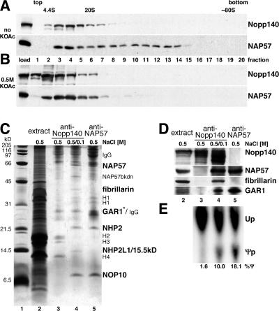
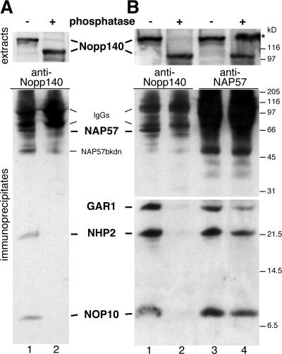
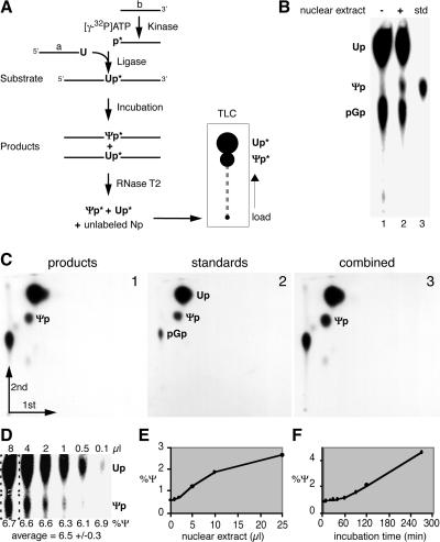
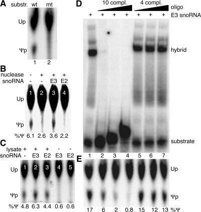
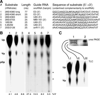
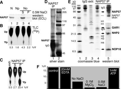
The isomerization of up to 100 uridines to pseudouridines (Psis) in eukaryotic rRNA is guided by a similar number of box H/ACA small nucleolar RNAs (snoRNAs), each forming a unique small nucleolar ribonucleoprotein particle (snoRNP) with the same four core proteins, NAP57 (also known as dyskerin or Cbf5p), GAR1, NHP2, and NOP10. Additionally, the nucleolar and Cajal body protein Nopp140 (Srp40p) associates with the snoRNPs. To understand the role of these factors in pseudouridylation, we established an in vitro assay system. Short site-specifically (32)P-labeled rRNA substrates were incubated with subcellular fractions, and the conversion of uridine to Psi was monitored by thin-layer chromatography after digestion to single nucleotides. Immunopurified box H/ACA core particles were sufficient for the reaction. SnoRNPs associated quantitatively and reversibly with Nopp140. However, pseudouridylation activity was independent of Nopp140, consistent with a chaperoning role for this highly phosphorylated protein. Although up to 14 bp between the snoRNA and rRNA were required for the in vitro reaction, rRNA pseudouridylation and release occurred in the absence of ATP and magnesium. These data suggest that substrate release takes place without RNA helicase activity but may be aided by the snoRNP core proteins.


Figures

FIG. 1.
FIG. 1.
Reversible association of Nopp140 with snoRNPs. Western blots of fractions separated on 10 to 30% glycerol gradients under physiological (A) or high-salt (B) conditions (0.5 M KOAc) and probed with anti-Nopp140 antibodies (upper panels) and anti-NAP57 antibodies (lower panels) are shown. The top and bottom of the gradient are indicated, as are the migrating positions of bovine serum albumin (4.4S) and thyroglobulin (20S). The 80S position was estimated by comparison of published gradients. (C) Silver-stained SDS-PAGE of immunoprecipitations from 0.5 M salt nucleolar extracts (lane 2) with anti-Nopp140 (lanes 3 and 4) and anti-NAP57 antibodies (lane 5). Before precipitation in lane 4, the extracts were diluted to 0.1 M salt. The specifically precipitating proteins (bold), the immunoglobulins (IgG), contaminating histones (H1 to H4), and a NAP57 breakdown product (NAP57bkdn) are indicated (see Fig. 6E for a detailed explanation). The asterisk draws attention to the fact that GAR1 is visible only in lane 5, not in lane 4, in which it comigrates with the anti-Nopp140 IgG light chains (see Fig. 1D and 6E) (53). Lane 2 contains 1/100 of the input used for the precipitations. (D) Western blots of the same samples as in panel C (lanes 2 to 5). Respective migration regions were probed with anti-Nopp140, -NAP57, -fibrillarin, and -GAR1 antibodies as indicated on the right-hand side and detected by enhanced chemiluminescence. (E) Autoradiograph of nucleotides separated by TLC after liberation from substrate 1, which was incubated with the same immunoprecipitates shown in panels C and D (lanes 3 to 5) to determine their pseudouridylase activity in vitro as described in the legend to Fig. 3.
FIG. 2.
FIG. 2.
Nopp140 dephosphorylation releases box H/ACA snoRNPs. Western blots of immunoprecipitates from high-salt nucleolar extracts diluted to 0.1 M salt (A) and from low-salt nuclear extracts (B) probed consecutively with antibodies against the antigens, indicated in bold between the panels, are shown. Prior to immunoprecipitations, the rat liver extracts were incubated in the absence (odd lanes) or presence (even lanes) of alkaline phosphatase. The dephosphorylation of Nopp140 in the extracts was documented by its mobility shift from 140 to 100 kDa (upper panels). The extracts were incubated with anti-Nopp140 antibodies (lanes 1 and 2) and anti-NAP57 antibodies (lane 3 and 4), and the coprecipitating antigens were visualized by Western blotting (lower panels). The lower half was exposed longer for ECL detection (B) (bottom panels). The asterisk refers to Nopp140 spillover from an adjacent lane on the gel, and additional labels are as described for Fig. 1.
FIG. 3.
FIG. 3.
In vitro assay for pseudouridylation of rRNA. (A) Schematic of the assay outlining the synthesis of the site-specifically labeled rRNA substrate by oligodeoxynucleotide-mediated ligation of the two independently synthesized 5′ (a) and 3′ (b) halves after labeling of b with 32P (p*). The reaction products are analyzed by nucleolytic liberation of the uridine (Up*) and Ψ 3′ monophosphates (Ψp*) and their separation by TLC followed by autoradiography. (B) Autoradiograph of a TLC demonstrating the production of Ψ only in the presence of HeLa nuclear extract (lane 2). Ψ 3′ monophosphate was loaded as standard (std, lane 3). Guanosine 5′,3′-diphosphate (pGp) was generated by the digest of incompletely ligated 3′-half substrate (b in panel A). (C) Analysis of the reaction products by two-dimensional TLC. The identity of the product Ψp (panel 1) is confirmed by the generation of a single spot when standard nucleotides (panel 2) are mixed with the product prior to chromatography (panel 3). (D) Quantitation by phosphorimager analysis varies minimally over an 80-fold difference in product loading on TLC. The amounts of Up and Ψp were each determined in rectangles (dotted lines) that had been background corrected, and the amount of Ψp was expressed as a percentage of total Up and Ψp (%Ψ). The production of Ψp increased with the amount of HeLa nuclear extract (E) and with increasing incubation time (F). Note that the higher activity in panel D than in panels E and F was caused by incubation in a different extract, BRL nucleolar extract.
FIG. 4.
FIG. 4.
In vitro pseudouridylation requires snoRNP-rRNA hybridization. (A) Autoradiograph of a TLC of uridine and Ψ liberated from the substrate after incubation with rat liver nucleolar extract. Ψ is produced from the wild-type rRNA substrate 1 (lane 1) but not a mutant substrate consisting of the 3′ half of substrate 1 but the 5′ half of substrate 3 (lane 2). (B) Pseudouridylation of substrate 1 in nuclear lysate pretreated with micrococcal nuclease (lanes 2 to 4) or not pretreated (lane 1). Addition of the cognate E3 snoRNA (lane 3) but not the noncomplementary E2 snoRNA (lane 4) partially restored the activity. Note that “trailing” of uridine often artificially elevated the numerical value of Ψ production under negative conditions (e.g., see lanes 2 and 4). (C) Ψ production in substrate 1 (lane 1) is enhanced in untreated nuclear lysates by E3 snoRNA (lane 2) but not E2 snoRNA (lane 3). Addition of either snoRNA alone failed to produce Ψ (lanes 4 and 5). (D) RNA-RNA gel shift analysis. Autoradiograph of a native polyacrylamide gel loaded with a mixture of the 32P-labeled rRNA substrate 2 and its cognate, unlabeled snoRNA E3 (lane 1). Addition of 10-fold-increasing concentrations of an unlabeled RNA oligonucleotide corresponding to the 5′ half of the rRNA substrate with 10 nt complementarity to E3 competed effectively for the gel shift (lanes 2 to 4), but addition of an oligonucleotide corresponding to the 3′ half with only 4-nt complementarity (lanes 5 to 7) did not. Note that the 5′-half oligonucleotide also had the ability to hybridize to and shift the substrate when present in sufficiently high concentrations (lanes 3 and 4). (E) Addition of equal amounts of substrate 2 to immunopurified snoRNPs (see Fig. 6) produced Ψ efficiently (lane 1). This activity was competed by the 5′-half oligonucleotide (lanes 2 to 4) but not the 3′-half oligonucleotide (lanes 5 to 7).
FIG. 5.
FIG. 5.
The assay works with multiple substrates and extracts. (A) The rRNA substrates used in this study are listed by number, the position of the uridine they represent in the respective human (1-6) and yeast (7) rRNAs, their length in nucleotides, the snoRNA guiding their pseudouridylation (specifying which of the two hairpins contains the complementarity to rRNA), and the nucleotide sequence with the target uridine (bold) and the complementary nucleotides (underlined). Note the replacement of cytidine 4381 by a guanosine in substrate 1 without an effect on activity. The sequences of the human rRNA are identical in rats, except that the numbering of the target uridines differs in substrates 1 and 2 (4141), 3 (4124), and 4 (3459). (B) Autoradiograph of the separated nucleotides liberated from substrates 1 to 7 after incubation in BRL nuclear lysates (1-3), rat liver nucleolar extracts (4-6), and yeast whole-cell extracts (7). Asterisks denote additional products that were detectable when incubations were performed in complex cell fractions and likely represented transphosphorylation products after substrate degradation. (C) Uridine is isomerized in the full-length substrate. After incubation in HeLa nuclear extract, substrate 2 was reisolated, analyzed by denaturing urea PAGE (top panel, lane 1), and compared to half of the substrate before incubation (lane 2). The gel was sectioned into three slices (white dotted boxes), and the RNA was eluted from each slice and digested for analysis of the single nucleotides by TLC (bottom panel).
FIG. 6.
FIG. 6.
Minimal requirements for in vitro pseudouridylation of rRNA. (A and B) Subfractionation of BRL nuclear lysate (lane 1) into nucleoplasm (lane 2) and 0.5 M NaCl nucleolar extract supernatant (lane 3) and pellet (lane 4) is shown. The fractions were analyzed by Western blotting with anti-NAP57 antibodies (A) and for their pseudouridylase activity towards substrate 1 by TLC (B). Note that the activity cofractionated with the putative pseudouridylase NAP57 in the nucleolar extract (lane 3). (C) The pseudouridylase activity was precipitated from rat liver nucleolar extracts (extr.) with anti-NAP57 antibodies (lane 3) and removed from the supernatant (lane 2). Fivefold less extract was assayed (lane 1) than was used in the immunoprecipitation experiments. (D) Analysis of the active NAP57 immunoprecipitate by SDS-9% PAGE and silver staining (lane 1). NAP57, the immunoglobulin heavy chains (IgG), and the molecular size markers (lane 2) are indicated. (E) Comparison on Coomassie blue-stained Tricine SDS-15% PAGE of the active NAP57 immunoprecipitate (lane 4) to nucleolar extract (lane 3) and anti-NAP57 antibodies (lane 2). Note that 3,000-fold more extract was used for the precipitation than was loaded in lane 3. The only specifically precipitated protein bands (arrows and dash), the immunoglobulin chains (IgG), the histones (H1 to 4), and the molecular size markers (lane 1) are indicated. The identity of the specifically precipitated bands was confirmed by consecutive probing of a Western blot with anti-GAR1, -NHP2, and -NOP10 antibodies (lane 5, lower half) and -NAP57 antibody (upper half). (F) The pseudouridylation of substrate 1 by the immunopurified snoRNPs bound to protein A-Sepharose was determined under the indicated conditions relative to the control reaction containing 2 mM MgCl2, 100 mM NaCl, 20 mM HEPES (pH 7.5), and 5% glycerol.

Similar articles

Cited by

References

    1. Aris, J., and G. Blobel. 1988. Identification and characterization of a yeast nucleolar protein that is similar to a rat liver nucleolar protein. J. Cell Biol. 107:17-31. - PMC - PubMed
    1. Bakin, A., and J. Ofengand. 1993. Four newly located pseudouridylate residues in Escherichia coli 23S ribosomal RNA are all at the peptidyltransferase center: analysis by the application of a new sequencing technique. Biochemistry 32:9754-9762. - PubMed
    1. Balakin, A. G., L. Smith, and M. J. Fournier. 1996. The RNA world of the nucleolus: two major families of small RNAs defined by different box elements with related functions. Cell 86:823-834. - PubMed
    1. Blobel, G., and V. Potter. 1966. Nuclei from rat liver: isolation method that combines purity with yield. Science 154:1662-1665. - PubMed
    1. Chang, M. S., H. Sasaki, M. S. Campbell, S. K. Kraeft, R. Sutherland, C. Y. Yang, Y. Liu, D. Auclair, L. Hao, H. Sonoda, L. H. Ferland, and L. B. Chen. 1999. HRad17 colocalizes with NHP2L1 in the nucleolus and redistributes after UV irradiation. J. Biol. Chem. 274:36544-36549. - PubMed

Publication types

MeSH terms