. 2002 Dec;22(24):8478-90.
doi: 10.1128/MCB.22.24.8478-8490.2002.

Expression of a novel, sterol-insensitive form of sterol regulatory element binding protein 2 (SREBP2) in male germ cells suggests important cell- and stage-specific functions for SREBP targets during spermatogenesis

Affiliations

Expression of a novel, sterol-insensitive form of sterol regulatory element binding protein 2 (SREBP2) in male germ cells suggests important cell- and stage-specific functions for SREBP targets during spermatogenesis

Hang Wang et al. Mol Cell Biol. 2002 Dec.

Abstract

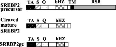
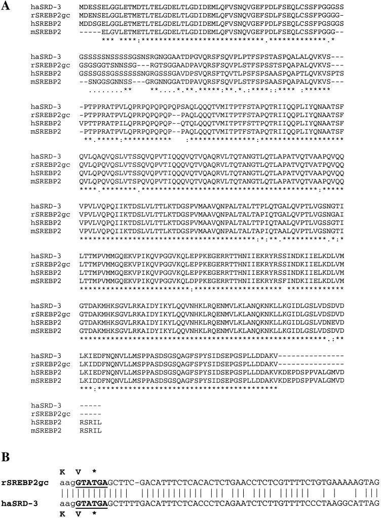
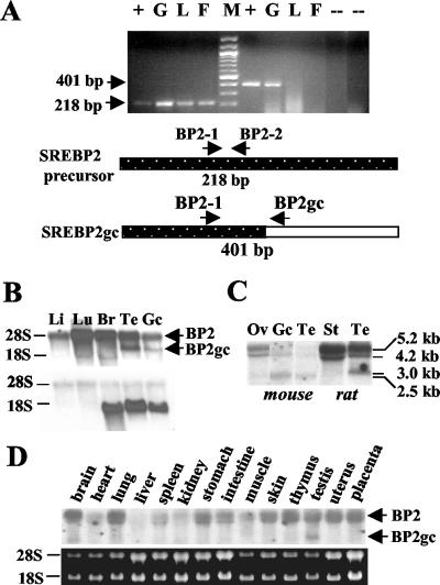
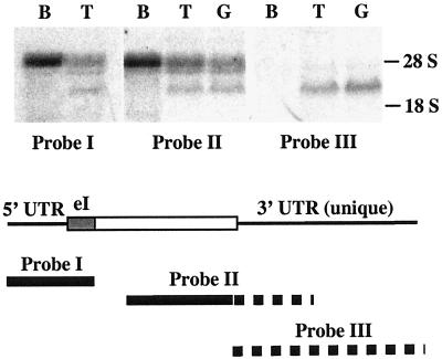
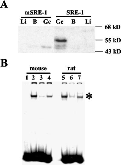
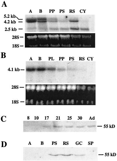
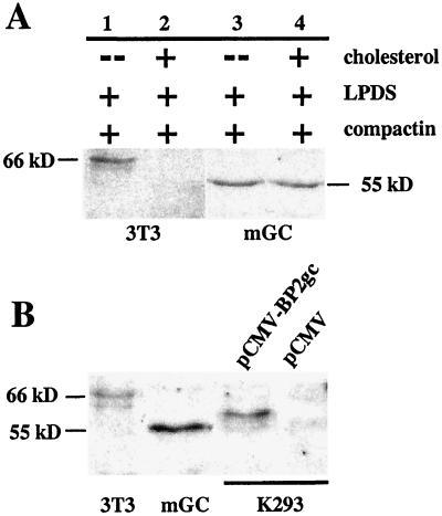
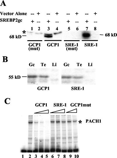
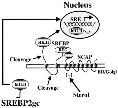
Cholesterol biosynthesis in somatic cells is controlled at the transcriptional level by a homeostatic feedback pathway involving sterol regulatory element binding proteins (SREBPs). These basic helix-loop-helix (bHLH)-Zip proteins are synthesized as membrane-bound precursors, which are cleaved to form a soluble, transcriptionally active mature SREBP that regulates the promoters for genes involved in lipid synthesis. Homeostasis is conferred by sterol feedback inhibition of this maturation process. Previous work has demonstrated the expression of SREBP target genes in the male germ line, several of which are highly up-regulated during specific developmental stages. However, the role of SREBPs in the control of sterol regulatory element-containing promoters during spermatogenesis has been unclear. In particular, expression of several of these genes in male germ cells appears to be insensitive to sterols, contrary to SREBP-dependent gene regulation in somatic cells. Here, we have characterized a novel isoform of the transcription factor SREBP2, which is highly enriched in rat and mouse spermatogenic cells. This protein, SREBP2gc, is expressed in a stage-dependent fashion as a soluble, constitutively active transcription factor that is not subject to feedback control by sterols. These findings likely explain the apparent sterol-insensitive expression of lipid synthesis genes during spermatogenesis. Expression of a sterol-independent, constitutively active SREBP2gc in the male germ line may have arisen as a means to regulate SREBP target genes in specific developmental stages. This may reflect unique roles for cholesterol synthesis and other functional targets of SREBPs during spermatogenesis.


Figures

FIG. 1.
FIG. 1.
Nucleotide and amino acid sequences of rat SREBP2gc cDNA. The numbers to the left refer to the positions of nucleotides and amino acids. The box denotes the conserved bHLH-Zip region, and the acidic N-terminal transactivation domain is underlined. The dashed lines indicate the primers used for 5′ and 3′ RACE-PCR. The dashed-lined boxes indicate potential germ cell-specific polyadenylation signals (52).
FIG. 2.
FIG. 2.
Schematic structures for different forms of SREBP2. Various domains are indicated as follows: acidic N-terminal transactivation domain (TA); serine- and glycine-rich (S) domain, as well as glutamine-rich (Q) domain; conserved bHLH-Zip DNA binding domain (bHZ); transmembrane domain (TM); and regulatory SCAP-binding domain (RSB). A 20-amino-acid sequence missing from the C terminus of SREBP2gc is also indicated by a small open box.
FIG. 3.
FIG. 3.
Comparison of sequences for rat SREBP2gc and SREBP2 from other species. (A) Protein sequences for SREBP2 from the mutant, sterol-resistant hamster cell line SRD-3 (haSRD-3) (53); rat SREBP2gc (rSREBP2gc); mature human SREBP2 (hSREBP2) (18); and the predicted mature form of mouse SREBP2 (mSREBP2), based on GenBank sequences (accession no. AAK54763) (Note that these sequences are incomplete at the N terminus.) Symbols below the amino acid sequences indicate the following relationships among different species: asterisks, identical amino acids; colons, high similarity; dots or open spaces, poor or no similarity, respectively. (B) Comparison of nucleotide sequences spanning the unique 3′-UTR, splice junction, and coding regions for rat SREBP2gc and SREBP2 from hamster SRD-3 cells. The 5′-splice sequence is underlined, and the stop codon is shown by an asterisk.
FIG. 4.
FIG. 4.
SREBP2 transcripts in rat and mouse tissues and male germ cells. (A) RT-PCR analysis. Two micrograms (each) of total RNA from adult rat spermatogenic cells (G), liver (L), and fat tissue (F) was analyzed. pBS-RBP2gc plasmid DNA was used as a positive control (+). No-RNA template and no-reverse transcriptase reactions served as negative controls (−). M, DNA size ladder. Shown are the 218-bp DNA fragment derived from SREBP2 mRNAs by using primers BP2-1 and BP2-2 (lanes to the left of size marker) and the 401-bp SREBP2gc-specific product generated with primers BP2-1 and BP2gc (lanes to the right of the size marker). (B, C, and D) Northern analysis of SREBP2 mRNAs with rat SREBP2gc cDNA as a probe. Twenty micrograms of total RNA was analyzed in each lane. (B) Rat liver (Li), lung (Lu), brain (Br), testis (Te), and enriched spermatogenic cells from adult testis (Gc). The same blot was probed for SREBP2gc (top) and rat proenkephalin (bottom) transcripts. RNA loading in the rat germ cell lane was reduced relative to rat testis, as shown by the lower signal for the germ cell-specific proenkephalin transcript (22) in this lane. (C) Mouse ovary (Ov), enriched adult mouse spermatogenic cells (Gc), mouse testis (Te), rat Sertoli cells (St), and rat testis (Te). (D) Analysis of multiple adult mouse tissues, as indicated. The results of ethidium bromide staining of 28S and 18S RNAs on the blot are shown below.
FIG. 5.
FIG. 5.
Identification of SREBP2gc RNA in rat spermatogenic cells. Twenty micrograms (each) of total RNA from rat brain (B), testis (T), and enriched adult rat spermatogenic cells (G) was examined. The positions of probes derived from the SREBP2gc cDNA are shown schematically below the Northern results. Solid lines show sequences common to the SREBP2p and SREBP2gc RNAs, while the dotted lines indicate sequences unique to the SREBP2gc transcript. eI, presumptive exon I region based on sequence homology with the hamster SREBP2 gene.
FIG. 6.
FIG. 6.
Analysis of SREBP2 mRNA translation and protein products in male germ cells. (A) Polysome analysis. Cytoplasmic RNA from mouse testis was fractionated in the presence of Mg2+ (HKM) or EDTA (HKE). Equal percentages of total RNA from fractions within each gradient were examined for SREBP2gc RNA by Northern analysis. RNA in lane 2 of the HKM gradient was underrepresented due to partial loss of sample. Gradient orientations are indicated below the Northern blot results, with larger polysomes localized to the denser (bottom) fractions. (B) Western analysis of SREBP2 protein in mouse and rat tissues and spermatogenic cells. (Left panel) Four micrograms (each) of microsomal protein from enriched adult mouse spermatogenic cells (G), brain (B), liver (Li), and lung (Lu) was examined. (Right panel) Analysis of nuclear extracts (5 μg) from enriched adult mouse spermatogenic cells (G), brain (B), kidney (K), and lung (Lu). (Lower panel) Five micrograms (each) of nuclear extract from enriched adult mouse and rat spermatogenic cells (G), mouse liver (Li), and rat lung (Lu) was examined. (C) Western blot of nuclear extracts (5 μg) from adult mouse spermatogenic cells (G) and sterol-depleted mouse 3T3-L1 cells (3T3). (D) Analysis of SREBP2gc protein in nuclei (Nu) and cytoplasmic (Cyt) fractions of adult mouse spermatogenic cells (5 μg per lane).
FIG. 7.
FIG. 7.
Detection of SRE-1 binding proteins in adult mouse spermatogenic cells and tissues. (A) Southwestern analysis of nuclear extracts (5 μg) from enriched spermatogenic cells from adult mouse testis (Gc), brain (B), and liver (Li). Wild-type (SRE-1) and mutant (mSRE-1) probes were used. (B) EMSA of SREBPs in mouse and rat spermatogenic cells. Nuclear extracts (1.5 μg) were examined for spermatogenic cells from adult mouse testis (lanes 2 to 4) and rat testis (lanes 5 to 7) by using the SRE-1 probe. Lanes: 1, probe only; 2 and 5, no competitor; 3 and 6, 10-fold excess of unlabeled SRE-1 wild-type competitor; 4 and 7, 10-fold excess of mutated SRE-1 competitor. The specific SREBP-SRE-1 complex is shown by an asterisk. The residual binding seen with the wild-type SRE-1 competitor was specifically eliminated at larger amounts of competitor (data not shown).
FIG. 8.
FIG. 8.
Developmental expression of SREBP2gc in male germ cells. (A) Northern analysis of purified mouse spermatogenic cells with rat SREBP2gc cDNA as a probe. Fifteen micrograms (each) of total RNA from spermatogonia type A (A), spermatogonia type B (B), prepubertal pachytene spermatocytes (PP), pachytene spermatocytes (PS), round spermatids (RS), and cytoplasts (Cy) was examined. Relative RNA loading is shown below by the ethidium bromide staining of the blot prior to hybridization. (B) Northern analysis of purified mouse spermatogenic cells with SREBP1c cDNA as a probe. Fractions are as in panel A, with the addition of preleptotene spermatocytes (PL), and the ethidium bromide-stained blot is shown below. (C) Western analysis of SREBP2gc protein in the developing mouse testis. Five micrograms (each) of testis nuclear extracts was examined from the indicated postnatal day mice and adults (Ad). (D) Western analysis of SREBP2gc protein in purified mouse spermatogenic cells. Five micrograms (each) of nuclear extracts from spermatogonia type A (A), spermatogonia type B (B), pachytene spermatocytes (PS), round spermatids (RS), enriched spermatogenic cells from adult mouse testis (Gc), and mouse sperm (SP) was examined.
FIG. 9.
FIG. 9.
Transcriptional activity of rat SREBP2gc. K293 cells were cotransfected with luciferase reporter plasmids for the LDLR promoter (A) or CYP51 promoter (B) and expression vectors for rat SREBP2gc (pCMV-BP2gc), constitutively active human SREBP2 (pCMV-BP2), or empty pCMV7 parent vector along with pCMV-β-galactosidase control plasmid. Data are normalized relative to β-galactosidase activity and are expressed as the fold increase in activity relative to the activity of the pCMV7 control plasmid. Shown are the means ± standard errors of three independent experiments.
FIG. 10.
FIG. 10.
Cell-type-specific regulation of SREBP2gc expression during spermatogenesis. (A) Effect of sterols on SREBP2 protein in mouse 3T3-L1 (3T3) and spermatogenic (mGC) cells. Lanes: 1 and 3, sterol-depleted culture conditions with compactin and LPDS alone; 2 and 4, sterol-loaded (plus cholesterol) conditions. (B) Size comparison for SREBP2 proteins from mouse 3T3-L1 cells (3T3), enriched spermatogenic cells from adult mouse testis (mGC), and K293 cells transfected with pCMV-BP2gc or pCMV empty vector. Five micrograms (each) of the nuclear extracts was examined by Western analysis, except for K293 cell samples (1.2 μg).
FIG. 11.
FIG. 11.
Comparative properties of SREBP2gc and PACH1. (A) Southwestern assay of bacterially expressed SREBP2gc protein. Bacterial extracts (7 μg) were assayed by using wild-type or mutant GCP1 or SRE-1 probes (shown below the lanes). The glutathione S-transferase (GST) fusion protein is predicted to be ∼68 kDa in size (29 kDa for GST plus 39 kDa for the SREBP2gc fragment). A nonspecific 70-kDa band (asterisk) was also detected with the GCP1 probe. (B) Southwestern analysis of SRE-1 and GCP1 binding proteins in rat spermatogenic cells and tissues. Five micrograms (each) of nuclear extracts from rat liver (Li), testis (Te), and enriched spermatogenic cells from adult rat testis (Gc) was used. (C) EMSA of GCP1 binding proteins in rat spermatogenic cells. One microgram of nuclear extracts of adult rat spermatogenic cells was examined with a GCP1 DNA probe. Competition experiments were performed with excess unlabeled GCP1, SRE-1, and mutated GCP1 DNAs. The PACH1 complex is shown by an asterisk.
FIG. 12.
FIG. 12.
Sterol-insensitive regulation of promoters in meiotic and early haploid spermatogenic cells by SREBP2gc. SREBP2gc bypasses the SCAP-dependent, sterol-regulated proteolytic processing pathway and directly enters the nucleus to regulate target genes in a constitutive manner.

Similar articles

Cited by

References

    1. Bajpai, M., G. Gupta, S. K. Jain, and B. S. Setty. 1998. Lipid metabolising enzymes in isolated rat testicular germ cells and changes associated with meiosis. Andrologia 30:311-315. - PubMed
    1. Beckman, J. K., and J. G. Coniglio. 1979. A comparative study of the lipid composition of isolated rat Sertoli and germinal cells. Lipids 14:262-267. - PubMed
    1. Bellve, A. R., J. C. Cavicchia, C. F. Millette, D. A. O'Brien, Y. M. Bhatnagar, and M. Dym. 1977. Spermatogenic cells of the prepubertal mouse. J. Cell Biol. 74:68-85. - PMC - PubMed
    1. Bellve, A. R., C. F. Millette, Y. M. Bhatnagar, and D. A. O'Brien. 1977. Dissociation of the mouse testis and characterization of isolated spermatogenic cells. J. Histochem. Cytochem. 25:480-494. - PubMed
    1. Blendy, J. A., K. H. Kaestner, G. F. Weinbauer, E. Nieschlag, and G. Schutz. 1996. Severe impairment of spermatogenesis in mice lacking the CREM gene. Nature 380:162-165. - PubMed

Publication types

MeSH terms