. 2002 Dec;22(24):8552-61.
doi: 10.1128/MCB.22.24.8552-8561.2002.

An ATR- and Chk1-dependent S checkpoint inhibits replicon initiation following UVC-induced DNA damage

Affiliations

An ATR- and Chk1-dependent S checkpoint inhibits replicon initiation following UVC-induced DNA damage

Timothy P Heffernan et al. Mol Cell Biol. 2002 Dec.

Abstract

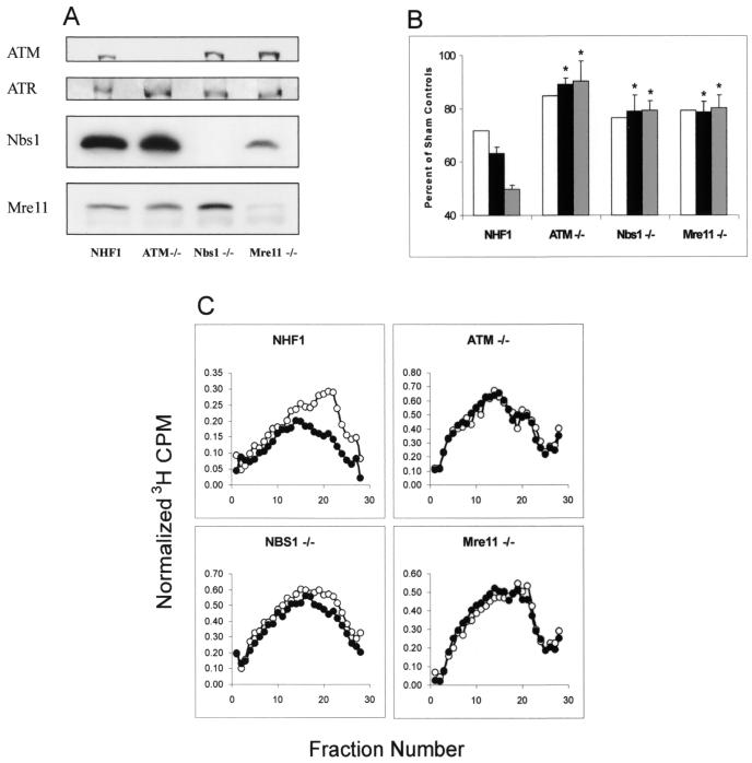
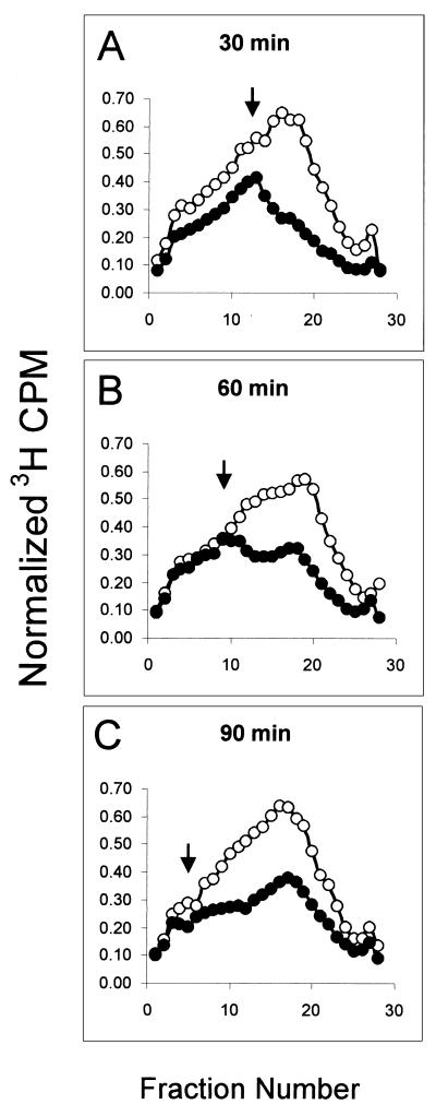
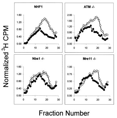
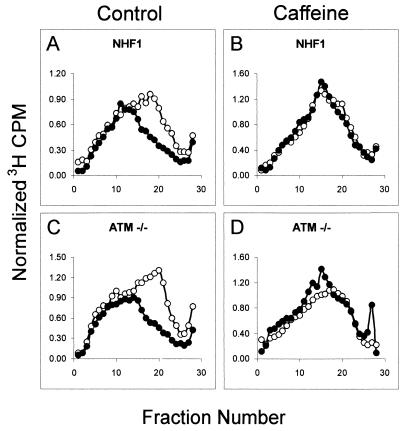
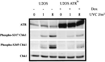
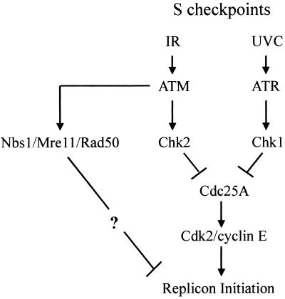
Inhibition of replicon initiation is a stereotypic DNA damage response mediated through S checkpoint mechanisms not yet fully understood. Studies were undertaken to elucidate the function of checkpoint proteins in the inhibition of replicon initiation following irradiation with 254 nm UV light (UVC) of diploid human fibroblasts immortalized by the ectopic expression of telomerase. Velocity sedimentation analysis of nascent DNA molecules revealed a 50% inhibition of replicon initiation when normal human fibroblasts were treated with a low dose of UVC (1 J/m(2)). Ataxia telangiectasia (AT), Nijmegen breakage syndrome (NBS), and AT-like disorder fibroblasts, which lack an S checkpoint response when exposed to ionizing radiation, responded normally when exposed to UVC and inhibited replicon initiation. Pretreatment of normal and AT fibroblasts with caffeine or UCN-01, inhibitors of ATR (AT mutated and Rad3 related) and Chk1, respectively, abolished the S checkpoint response to UVC. Moreover, overexpression of kinase-inactive ATR in U2OS cells severely attenuated UVC-induced Chk1 phosphorylation and reversed the UVC-induced inhibition of replicon initiation, as did overexpression of kinase-inactive Chk1. Taken together, these data suggest that the UVC-induced S checkpoint response of inhibition of replicon initiation is mediated by ATR signaling through Chk-1 and is independent of ATM, Nbs1, and Mre11.


Figures

FIG. 1.
FIG. 1.
Telomerase-expressing fibroblasts from patients with familial cancer syndromes display the RDS phenotype. (A) Protein extracts were prepared from NHF1, ATM−/−, Nbs1−/−, and Mre11−/− fibroblasts, and 100 μg of total protein was analyzed by western immunoblot analysis. (B) RDS results. Cells were grown in the presence of [14C]thymidine for ∼40 h to label DNA uniformly and then in nonradioactive medium overnight. Cells were sham treated or exposed to gamma rays, incubated at 37°C for 30 min, and then labeled for 15 min in medium containing [3H]thymidine. Net 3H radioactivity corrected for 14C spillover was normalized to cell number (total 14C radioactivity). The normalized 3H counts per minute was graphed as a percent of sham controls (n = 3). White bar, 2.5 Gy; black bar, 5.0 Gy; gray bar, 7.5 Gy. *, significant difference from NHF1 (P < 0.01). (C) Cells were uniformly labeled as described above and either sham treated or irradiated with 1.5 Gy IR. Following irradiation, cells were incubated for 30 min at 37°C and then pulse-labeled with [3H]thymidine for 15 min. Cells were harvested, and nascent DNA was separated by velocity sedimentation (see Materials and Methods). Net 3H radioactivity corrected for 14C spillover was normalized to cell number (total 14C radioactivity). ○, nascent DNA distributions from sham-treated cells; •, distributions from cultures irradiated with 1.5 Gy.
FIG. 2.
FIG. 2.
UVC-induced DNA damage inhibits replicon initiation. NHF1 cells were uniformly labeled as described above and sham treated or irradiated with 1 J/m2 UVC. Following treatment, cells were incubated in reserved medium for either 30 (A), 60 (B), or 90 (C) min prior to a 15-min pulse with [3H]thymidine. Velocity sedimentation analysis of nascent DNA was done as described in the legend to Fig. 1C. Open circles represent profiles from sham-treated cells, while closed circles represent those from UVC-irradiated cultures. Arrows point to the position in which the nascent DNA distribution from irradiated cells diverges noticeably from that observed with sham-treated cells.
FIG. 3.
FIG. 3.
UVC-induced inhibition of replicon initiation is independent of ATM, Nbs1, and Mre11. Indicated cell lines were uniformly labeled and sham treated or irradiated with 1 J/m2 UVC. Nascent DNA distribution profiles were determined as described in the legend to Fig. 1C. Cultures were incubated for 30 min and then pulse-labeled with [3H]thymidine. ○, sham-treated cells; •, UVC-irradiated cells. The average inhibition of replicon initiation in cells exposed to 1 J/m2 UVC was 50% (n = 19; range, 39 to 57%) in NHF1; 49% (n = 7) in ATM−/−; 40% (n = 2) in NBS1−/−; and 42% (n = 2) in Mre11−/− cells.
FIG. 4.
FIG. 4.
UVC-induced inhibition of replicon initiation is sensitive to caffeine. NHF1 and ATM−/− cells were uniformly labeled as described above and incubated for 30 min in medium alone (A and C) or medium containing 3 mM caffeine (B and D) for 30 min. Cells were sham treated or irradiated with 1 J/m2 UVC, incubated in reserved medium for 30 min, and then pulse-labeled with [3H]thymidine for 15 min. Cells treated with caffeine remained in the presence of this inhibitor throughout the treatment period. Cells were harvested, and nascent DNA was separated by velocity sedimentation as described in the legend to Fig. 1C. ○, sham-treated cells; •, UVC-irradiated cells.
FIG. 5.
FIG. 5.
The UVC-induced S checkpoint is ATR dependent. (A) U2OS cells were grown in medium containing 1 μg of doxycycline (Dox)/ml for 48 h to induce the expression of either ATRwt or ATRki. Protein extracts were prepared, and 100 μg of extract was analyzed by Western immunoblot analysis. (B) U2OS cells were uniformly labeled in the absence (uninduced) or the presence (48 h) of 1 μg of doxycycline/ml and pretreated with either medium or 2 mM caffeine 30 min prior to a sham treatment or irradiation with 1 J/m2 UVC. Following irradiation, cells were incubated for 30 min at 37°C and then pulse-labeled with [3H]thymidine for 15 min in their respective medium (with or without doxycycline; with or without caffeine). Cells were harvested, and nascent DNA was separated by velocity sedimentation as described in the legend to Fig. 1C. ○, sham-treated cells; •, UVC-irradiated cells. (C) The degree of inhibition of replicon initiation was calculated from velocity sedimentation profiles of separate experiments and graphed as the average percentage of control (n = 6). *, significant difference from uninduced controls (P < 0.0001).
FIG. 6.
FIG. 6.
ATR-dependent Chk1 phosphorylation following UVC-induced DNA damage. U2OS cells were grown for 48 h in the presence or absence of 1 μg of doxycycline (Dox)/ml to induce the expression of ATRki. Cells were either sham treated (0) or irradiated with 1 or 8 J/m2 UVC. Cells were incubated for 30 min in reserved medium and then harvested. Protein extracts were prepared, and 100 μg of extract was analyzed by Western immunoblot analysis.
FIG. 7.
FIG. 7.
Chk1 activation is required for the UVC-induced S checkpoint. (A) NHF1 and ATM−/− cells were uniformly labeled as described and incubated in medium containing dimethyl sulfoxide (A and C) or 100 nM UCN-01 (B and D) for 30 min. Cells were either sham treated or irradiated with 1 J/m2 UVC and incubated in reserved medium for 30 min prior to a 15-min pulse with [3H]thymidine in their respective media. Cells were harvested, and nascent DNA was separated by velocity sedimentation as described in the legend to Fig. 1C. ○, sham-treated cells; •, UVC-irradiated cells. (E) U2OS cells were infected with adenovirus encoding Chk1wt, Chk1ki, or GFP at an MOI of 50 for 15 h. Protein extracts were prepared, and Chk1 expression was measured by Western immunoblot analysis. (F) U2OS cells were grown in the presence of [14C]thymidine for ∼40 h to label DNA uniformly and then in nonradioactive medium containing adenovirus at an MOI of 50 for 15 h. Cells were either sham treated (black bars) or exposed to 1 J/m2 UVC (white bars), incubated for 30 min at 37°C, and then labeled for 15 min in medium containing [3H]thymidine. Net 3H radioactivity corrected for 14C spillover was normalized to cell number (total 14C radioactivity). The normalized 3H counts per minute was graphed as a percentage of the sham control value (n = 4 to 7). *, significant difference from sham controls (P < 0.0001).
FIG. 8.
FIG. 8.
Schematic model of ATM- and ATR-dependent S checkpoint signaling pathways.

References

    1. Abraham, R. T. 2001. Cell cycle checkpoint signaling through the ATM and ATR kinases. Genes Dev. 15:2177-2196. - PubMed
    1. Adler, V., S. Y. Fuchs, J. Kim, A. Kraft, M. P. King, J. Pelling, and Z. Ronai. 1995. jun-NH2-terminal kinase activation mediated by UV-induced DNA lesions in melanoma and fibroblast cells. Cell Growth Differ. 6:1437-1446. - PubMed
    1. Banin, S., L. Moyal, S. Shieh, Y. Taya, C. W. Anderson, L. Chessa, N. I. Smorodinsky, C. Prives, Y. Reiss, Y. Shiloh, and Y. Ziv. 1998. Enhanced phosphorylation of p53 by ATM in response to DNA damage. Science 281:1674-1677. - PubMed
    1. Blasina, A., B. D. Price, G. A. Turenne, and C. H. McGowan. 1999. Caffeine inhibits the checkpoint kinase ATM. Curr. Biol. 9:1135-1138. - PubMed
    1. Boyer, J. C., W. K. Kaufmann, B. P. Brylawski, and M. Cordeiro-Stone. 1990. Defective postreplication repair in xeroderma pigmentosum variant fibroblasts. Cancer Res. 50:2593-2598. - PubMed

Publication types

MeSH terms

Substances