. 2002 Dec;22(24):8580-91.
doi: 10.1128/MCB.22.24.8580-8591.2002.

Essential, nonredundant role for the phosphoinositide 3-kinase p110delta in signaling by the B-cell receptor complex

Affiliations

Essential, nonredundant role for the phosphoinositide 3-kinase p110delta in signaling by the B-cell receptor complex

Shiann-Tarng Jou et al. Mol Cell Biol. 2002 Dec.

Abstract

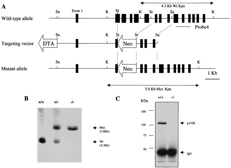
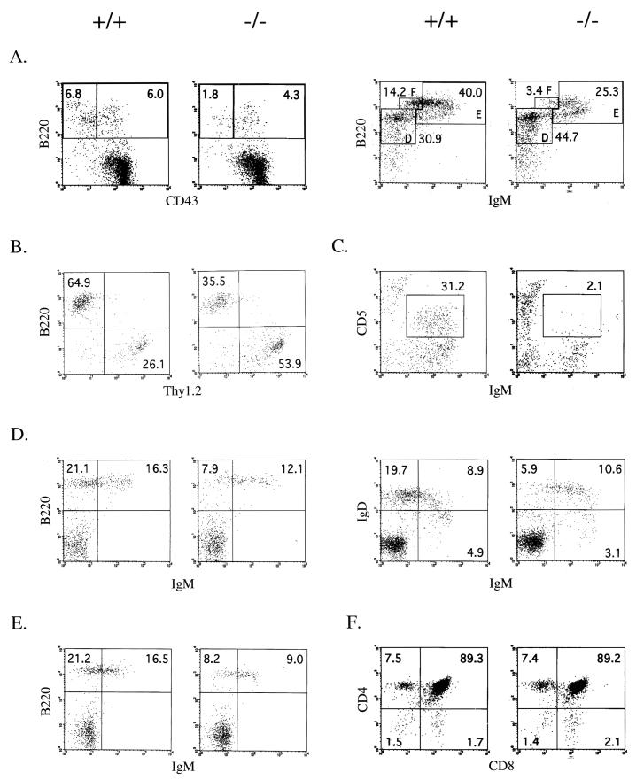
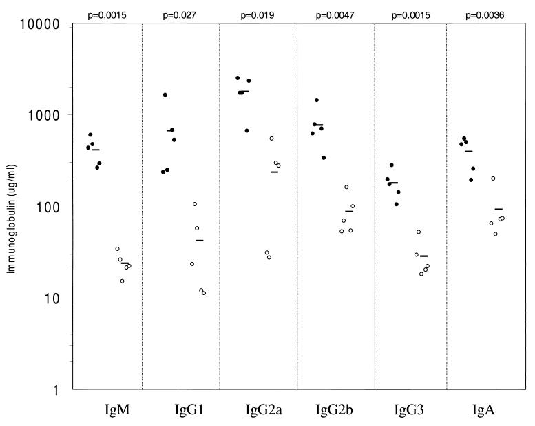
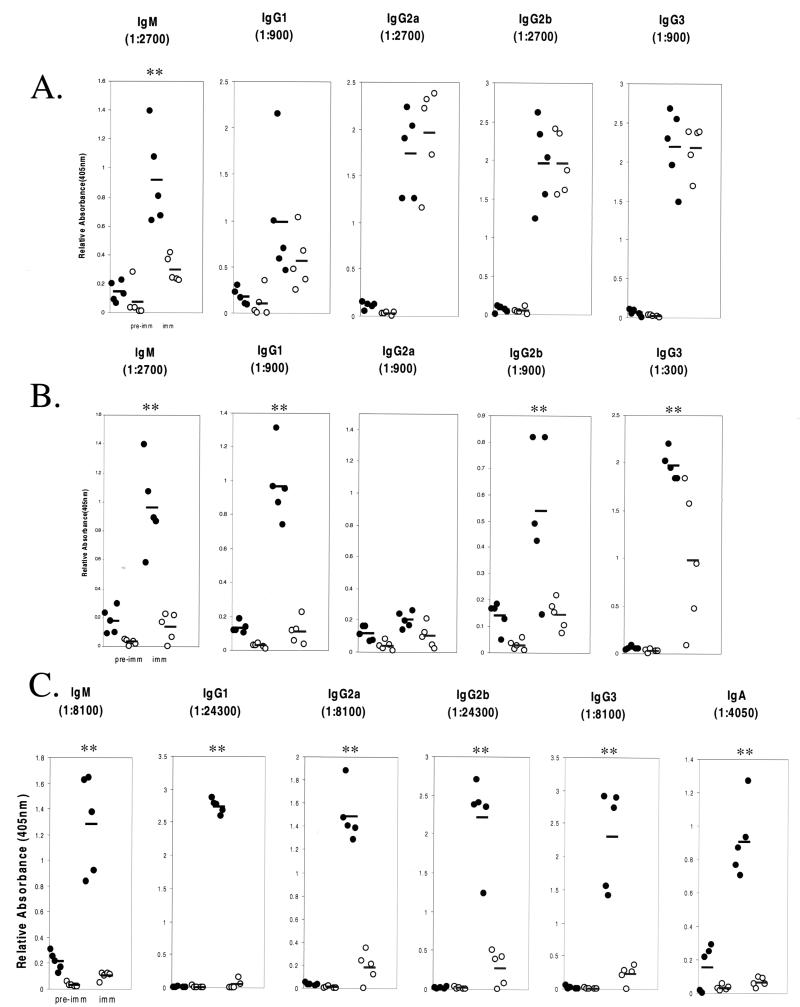
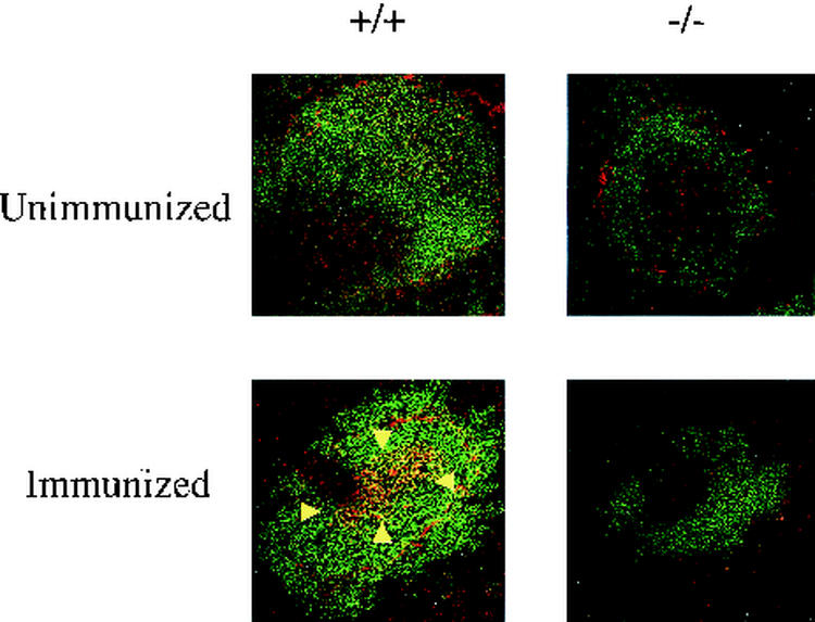
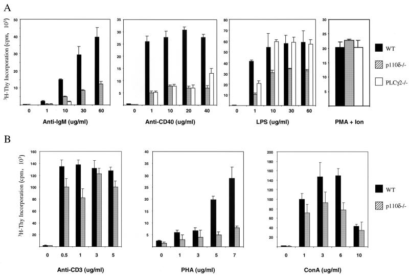
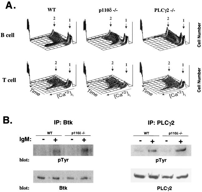
Many receptor and nonreceptor tyrosine kinases activate phosphoinositide 3-kinases (PI3Ks). To assess the role of the delta isoform of the p110 catalytic subunit of PI3Ks, we derived enzyme-deficient mice. The mice are viable but have decreased numbers of mature B cells, a block in pro-B-cell differentiation, and a B1 B-cell deficiency. Both immunoglobulin M receptor-induced Ca(2+) flux and proliferation in response to B-cell mitogens are attenuated. Immunoglobulin levels are decreased substantially. The ability to respond to T-cell-independent antigens is markedly reduced, and the ability to respond to T-cell-dependent antigens is completely eliminated. Germinal center formation in the spleen in response to antigen stimulation is disrupted. These results define a nonredundant signaling pathway(s) utilizing the delta isoform of p110 PI3K for the development and function of B cells.


Figures

FIG. 1.
FIG. 1.
Targeted disruption of murine p110δ gene. (A) A targeting construct that would replace the indicated exons with a neomycin resistance cassette was developed. The locations of the 3′ external probe and PCR primers (P1, P2, and P3) for genotyping are indicated. Restriction enzyme sites: Sa, SacI; K, KpnI; St, StuI. DTA, diphtheria toxin A; Wt, wild type. (B) Southern blot analysis of KpnI-digested genomic DNA from p110δ heterozygous mice probed with the indicated probe. The positions of the mutant 5.8-kb and wild-type 4.3-kb KpnI fragments are indicated. (C) Absence of p110δ protein expression in p110δ−/− mice. Total splenic lysate (2 mg) was used for immunoprecipitation and Western blot analysis with rabbit polyclonal anti-p110δ antibodies (see Materials and Methods).
FIG. 2.
FIG. 2.
Flow cytometric analysis of B- and T-cell development in the primary and secondary lymphoid organs. Total cells isolated from 3- to 4-month-old mice were stained with the indicated antibodies and analyzed by FACS. The numbers in each region indicate the percentage of the total cells plotted. The data shown are representative of those from a minimum of six pairs of mice examined. (A) Early and late stages (left and right, respectively) of B-cell maturation in the bone marrow. Left panel, B220 versus CD43 profiles of IgM-gated cells; right panels, B220 versus IgM profiles of CD43-gated cells (fractions D, E, and F). (B) Analysis of splenic B and T cells, showing profile of B220 versus Thy1.2 gated lymphocytes. (C) Analysis of peritoneal B1a cells, displayed as the profile of IgM+ CD5+ cells within the lymphocyte gate. (D) Maturation stages of splenic B cells. Left panels, B220 versus IgM profiles of gated lymphocytes; right panels, IgD versus IgM profiles of gated lymphocytes. (E) Analysis of lymph node B cells, showing B220 IgM profiles of gated lymphocytes. (F) Maturation of thymocytes is unimpaired (CD4 versus CD8 profile of gated thymocytes).
FIG. 3.
FIG. 3.
Decreased serum immunoglobulin concentrations in p110δ−/− mice. Levels of immunoglobulin isotypes in serum in unimmunized wild type or p110δ-deficient mice were determined by ELISA. The values for each individual mouse tested are plotted on a logarithmic graph. Mean values are indicated by boldface lines. P values as determined by Student's t test are indicated.
FIG. 4.
FIG. 4.
Defective humoral response to thymus-independent antigens and lack of a humoral response to a thymus-dependent antigen in p110δ-deficient mice. Wild-type (filled circles) or p110δ-deficient (open circles) mice were immunized with either TNP-LPS, a TI-I antigen (A); TNP-Ficoll, a TI-II antigen (B); or TNP-KLH, a T-dependent antigen (C). The serum TNP-specific immunoglobulin isotype response was measured by ELISA. The relative absorbance values at 405 nm for the indicated dilution are plotted for each individual animal. Mean values are indicated by boldface lines. Statistically significant differences (P < 0.05) are indicated (**).
FIG. 5.
FIG. 5.
Germinal center formation in TNP-KLH-stimulated 4-month-old mice. Germinal centers (arrowheads) were stained with biotinylated PNA followed by streptavidin-tetramethyl rhodamine isocyanate (red). B cells were stained with FITC-B220 (green).
FIG. 6.
FIG. 6.
Impaired in vitro proliferative response of p110δ-deficient lymphocytes. (A). Purified splenic B cells from 3- to 4-month-old wild-type mice (WT), p110δ−/− mice, and PLCγ2−/− mice were incubated in medium alone or in medium containing the indicated concentrations of various stimuli. Proliferative responses are expressed as mean counts of triplicates from a single [3H]thymidine incorporation assay; error bars indicate standard deviations. Results are representative of those from four to six experiments. Ion, ionomycin. (B) Splenic T cells were incubated in medium alone or in medium containing the indicated concentrations of various stimuli. Proliferative responses are expressed as mean counts of triplicates from a single [3H]thymidine incorporation assay; error bars indicate standard deviations. Results are representative of those from four to six experiments.
FIG. 7.
FIG. 7.
Induction of Ca2+ mobilization and tyrosine phosphorylation in p110δ-deficient mice. (A) Induction of Ca2+ was measured by flow cytometry following stimulation of Indo-1-labeled splenic B cells with anti-IgM or of splenic T cells with anti-CD3 followed by anti-IgG. Arrow 1, time point of specific stimulation; arrow 2, time point of ionomycin addition. The results are representative of those from four independent experiments. WT, wild type. (B) Purified splenic B cells from wild-type and p110δ−/− mice were stimulated with anti-IgM (20 μg/ml) for 3 min and analyzed for the induction of tyrosine phosphorylation (pTyr) of Btk and PLCγ2. IP, immunoprecipitation.

Similar articles

Cited by

References

    1. Bi, L., I. Okabe, D. J. Bernard, and R. L. Nussbaum. 2002. Early embryonic lethality in mice deficient in the p110beta catalytic subunit of PI 3-kinase. Mamm. Genome 13:169-172. - PubMed
    1. Bi, L., I. Okabe, D. J. Bernard, A. Wynshaw-Boris, and R. L. Nussbaum. 1999. Proliferative defect and embryonic lethality in mice homozygous for a deletion in the p110alpha subunit of phosphoinositide 3-kinase. J. Biol. Chem. 274:10963-10968. - PubMed
    1. Buhl, A. M., C. M. Pleiman, R. C. Rickert, and J. C. Cambier. 1997. Qualitative regulation of B cell antigen receptor signaling by CD19: selective requirement for PI3-kinase activation, inositol-1,4,5-trisphosphate production and Ca2+ mobilization. J. Exp. Med. 186:1897-1910. - PMC - PubMed
    1. Chen, W. S., P. Z. Xu, K. Gottlob, M. L. Chen, K. Sokol, T. Shiyanova, I. Roninson, W. Weng, R. Suzuki, K. Tobe, T. Kadowaki, and N. Hay. 2001. Growth retardation and increased apoptosis in mice with homozygous disruption of the Akt1 gene. Genes Dev. 15:2203-2208. - PMC - PubMed
    1. Cho, H., J. Mu, J. K. Kim, J. L. Thorvaldsen, Q. Chu, E. B. Crenshaw III, K. H. Kaestner, M. S. Bartolomei, G. I. Shulman, and M. J. Birnbaum. 2001. Insulin resistance and a diabetes mellitus-like syndrome in mice lacking the protein kinase Akt2 (PKB beta). Science 292:1728-1731. - PubMed

Publication types

MeSH terms

LinkOut - more resources