. 2002 Dec;22(24):8681-94.
doi: 10.1128/MCB.22.24.8681-8694.2002.

Downregulation of c-Jun expression by transcription factor C/EBPalpha is critical for granulocytic lineage commitment

Affiliations

Downregulation of c-Jun expression by transcription factor C/EBPalpha is critical for granulocytic lineage commitment

Janki Rangatia et al. Mol Cell Biol. 2002 Dec.

Abstract

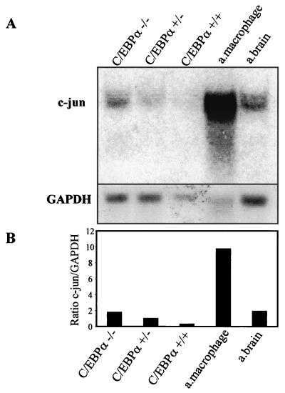
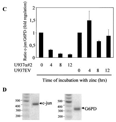
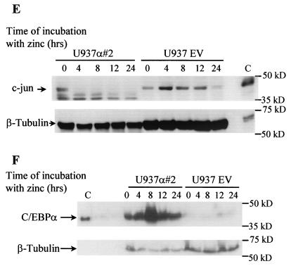
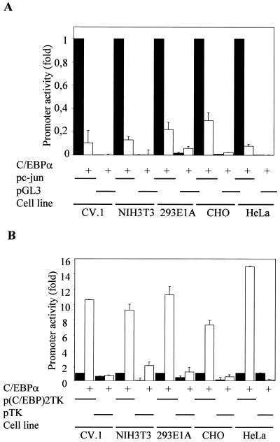
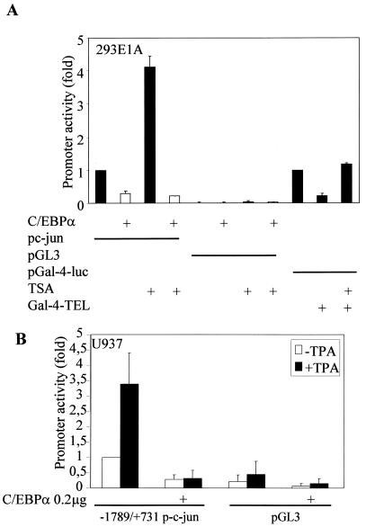
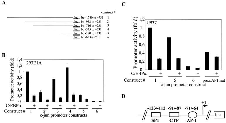
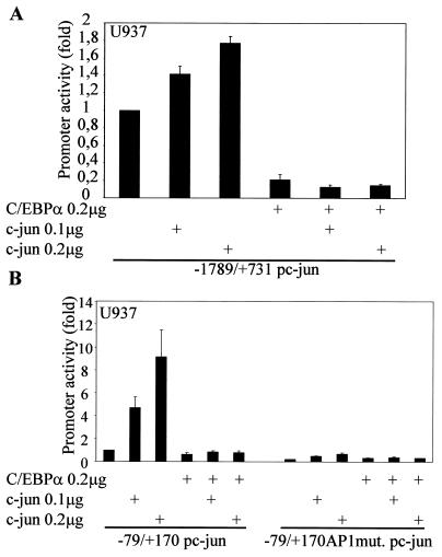
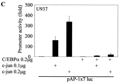
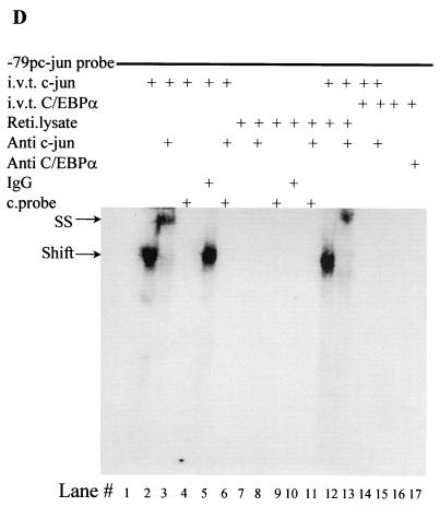
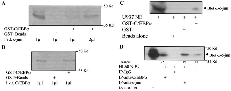
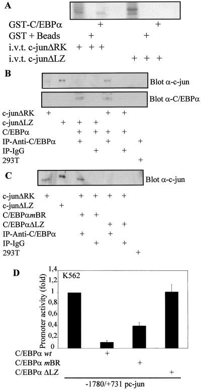
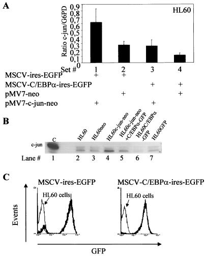
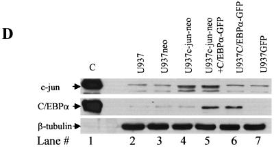
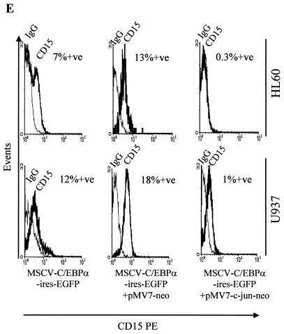
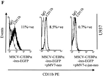
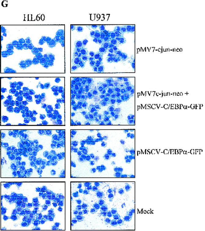
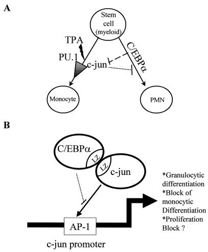
The transcription factor C/EBPalpha is crucial for the differentiation of granulocytes. Conditional expression of C/EBPalpha triggers neutrophilic differentiation, and C/EBPalpha can block 12-O-tetradecanoylphorbol-13-acetate-induced monocytic differentiation of bipotential myeloid cells. In C/EBPalpha knockout mice, no mature granulocytes are present. A dramatic increase of c-Jun mRNA in C/EBPalpha knockout mouse fetal liver was observed. c-Jun, a component of the AP-1 transcription factor complex and a coactivator of the transcription factor PU.1, is important for monocytic differentiation. Here we report that C/EBPalpha downregulates c-Jun expression to drive granulocytic differentiation. An ectopic increase of C/EBPalpha expression decreases the c-Jun mRNA level, and the human c-Jun promoter activity is downregulated eightfold in the presence of C/EBPalpha. C/EBPalpha and c-Jun interact through their leucine zipper domains, and this interaction prevents c-Jun from binding to DNA. This results in downregulation of c-Jun's capacity to autoregulate its own promoter through the proximal AP-1 site. Overexpression of c-Jun prevents C/EBPalpha-induced granulocytic differentiation. Thus, we propose a model in which C/EBPalpha needs to downregulate c-Jun expression and transactivation capacity for promoting granulocytic differentiation.


Figures

FIG. 1.
FIG. 1.
Reciprocal expression of C/EBPα and c-Jun. (A) Northern blot analysis showing the expression of c-Jun in day 19 fetal livers from C/EBPα+/+, C/EBPα+/−, and C/EBPα−/− mice, adult (a.) mouse brain, and peritoneal macrophages. Total RNA (10 μg) for each sample was electrophoresed, transferred, and hybridized with an α-32P-labeled c-Jun 1.1-kb BamHI-EcoRI cDNA fragment and a G6PD control fragment. (B) Ratio of c-Jun to G6PD from the Northern analysis data in panel A. (C) The U937 α#2 and U937 EV cell lines were induced with 100 μM zinc sulfate, and total RNA was collected at 0, 4, 8, 12, and 16 h. cDNA from 1 μg of total RNA was used for real-time PCR using c-Jun- and G6PD-specific primers. The error bars represent standard errors of the means from three independent experiments. (D) Specific real-time PCR products of c-Jun and G6PD as observed after 1.2% agarose gel electrophoresis. Numbers on the left of each panel are molecular sizes in kilodaltons. (E) Western blot analysis showing the expression of c-Jun protein from whole-cell extracts of the U937 α#2 and U937 EV cell lines after 0, 4, 8, 12, and 24 h of zinc induction. Immunodetection was performed using c-Jun specific antibody. Lane C, in vitro-translated c-Jun positive control. β-Tubulin expression from the same blot is shown as a loading control. (F) Western blot analysis of the whole-cell extracts from panel E for C/EBPα expression with specific antibody. Lane C, in vitro-translated C/EBPα and loading control for the same Western blot.
FIG. 1.
FIG. 1.
Reciprocal expression of C/EBPα and c-Jun. (A) Northern blot analysis showing the expression of c-Jun in day 19 fetal livers from C/EBPα+/+, C/EBPα+/−, and C/EBPα−/− mice, adult (a.) mouse brain, and peritoneal macrophages. Total RNA (10 μg) for each sample was electrophoresed, transferred, and hybridized with an α-32P-labeled c-Jun 1.1-kb BamHI-EcoRI cDNA fragment and a G6PD control fragment. (B) Ratio of c-Jun to G6PD from the Northern analysis data in panel A. (C) The U937 α#2 and U937 EV cell lines were induced with 100 μM zinc sulfate, and total RNA was collected at 0, 4, 8, 12, and 16 h. cDNA from 1 μg of total RNA was used for real-time PCR using c-Jun- and G6PD-specific primers. The error bars represent standard errors of the means from three independent experiments. (D) Specific real-time PCR products of c-Jun and G6PD as observed after 1.2% agarose gel electrophoresis. Numbers on the left of each panel are molecular sizes in kilodaltons. (E) Western blot analysis showing the expression of c-Jun protein from whole-cell extracts of the U937 α#2 and U937 EV cell lines after 0, 4, 8, 12, and 24 h of zinc induction. Immunodetection was performed using c-Jun specific antibody. Lane C, in vitro-translated c-Jun positive control. β-Tubulin expression from the same blot is shown as a loading control. (F) Western blot analysis of the whole-cell extracts from panel E for C/EBPα expression with specific antibody. Lane C, in vitro-translated C/EBPα and loading control for the same Western blot.
FIG. 2.
FIG. 2.
C/EBPα downregulates the c-Jun promoter activity. (A) Transient cotransfection of a c-Jun promoter reporter construct (bp −1780 to +731) and pGL3 with or without C/EBPα in CV.1, NIH 3T3, 293E1A, CHO, and HeLa cells. Solid bars, values for promoter alone; open bars, cotransfection with C/EBPα. The pRL-0 Renilla luciferase construct was cotransfected to normalize for transfection efficiency. Error bars indicate standard errors of the means. (B) Effect of transient cotransfection of C/EBPα on the positive control p(C/EBP)2TK-luciferase reporter construct, indicating the transactivation capacity of C/EBPα in these cell lines. The pTK-luciferase reporter construct served as a negative control. Solid bars, values for promoter alone; open bars, fold promoter activities in the presence of C/EBPα.
FIG. 3.
FIG. 3.
C/EBPα does not recruit a TSA-sensitive corepressor complex and blocks TPA-induced c-Jun promoter activity. (A) Transient-cotransfection experiments in the 293E1A cell line with the c-Jun promoter construct and C/EBPα in the presence or absence of TSA (100 nM). pGal-4-luc with Gal-4-TEL and TSA was used as a positive control for functionally active TSA. (B) U937 cells (106 in six-well plates) were transfected with 0.55 μg of c-Jun promoter construct (bp −1780 to +731) or pGL3, with or without 0.4 μg of C/EBPα expression plasmid or empty vector, and 0.05 μg of pRL-0. The cells were transfected by using the Effectene protocol (Qiagen). At 12 h posttransfection, TPA (100 nM) was added to the respective wells and further incubated at 37°C for 24 to 30 h. The pRL-0 Renilla luciferase construct was cotransfected to normalize for transfection efficiency. The results are the means from three independent experiments, and error bars represent the standard errors of mean values for each set.
FIG. 4.
FIG. 4.
c-Jun promoter mapping to identify the region important for C/EBPα-mediated downregulation. (A) Schematic presentation of various c-Jun promoter 5′ deletion constructs used for the transient-transfection experiment. (B) 293E1A cells (104 per well in 24-well plates) were transfected with 0.25 μg of 5′ c-Jun promoter deletion constructs bp −1780/+731, bp −953/+731, bp −716/+731, bp −345/+731, bp −180/+731, and bp −63/+731 with or without 0.2 μg of C/EBPα or empty vector and 0.05 μg of pRL-0. (C) U937 cells (106 per well in six-well plates) were transfected with 0.55 μg of 5′ c-Jun promoter deletion constructs bp −1780/+731, bp −180/+731, bp −63/+731, and bp −1780/+731 proximal AP-1 mutant c-Jun promoter with or without C/EBPα expression plasmid or empty vector and 0.05 μg of pRL-0. The cells were transfected by using the Effectene protocol. The pRL-0 Renilla luciferase construct was cotransfected to normalize for transfection efficiency. The results are the means from three independent experiments, and error bars represent the standard errors of mean values for each set. (D) Schematic presentation of the c-Jun promoter region between bp −180 and −63. This region contains a proximal AP-1 site, CTF site, and SP-1 site.
FIG. 5.
FIG. 5.
C/EBPα blocks the autoregulatory capacity of c-Jun by preventing c-Jun from binding to the proximal AP-1 site in the c-Jun promoter. (A) Transient transfections in U937 myeloid cells were performed using a bp −1780/+731 c-Jun promoter construct, 0.2 μg of C/EBPα expression plasmid, and increasing amounts of c-Jun expression plasmid (0.1 and 0.2 μg). Error bars indicate standard errors of the means. (B) bp −79/+190 and bp −79/+190 mutated AP-1 site c-Jun promoter constructs were transiently transfected with 0.2 μg of C/EBPα expression plasmid and increasing concentrations of c-Jun expression plasmid. (C) The AP-1 luciferase construct containing seven repeats of an AP-1 binding site was used for transient transfection with c-Jun and C/EBPα in U937 myeloid cells. (D) Electrophoretic mobility shift assay using [γ-32P]ATP-labeled bp −82/−53 c-Jun promoter oligonucleotide spanning the proximal AP-1 site was performed using in vitro-translated (i.v.t.) c-Jun (lanes 2 to 6 and 12 to 15) and C/EBPα (lanes 12, 13, 16, and 17) proteins, rabbit reticulocyte (Reti.) lysate (lanes 7 to 11, 14, and 15), c-Jun-specific antibody (lanes 3, 6, 8, 11, 13, and 15), normal rabbit IgG (lanes 4 and 9), C/EBPα-specific antibody (lane 17), and self unlabeled competitor probe (lanes 5, 6, 10, and 11). Arrows show the c-Jun shifted band (Shift) and the supershifted higher band with c-Jun-specific antibody (SS).
FIG. 5.
FIG. 5.
C/EBPα blocks the autoregulatory capacity of c-Jun by preventing c-Jun from binding to the proximal AP-1 site in the c-Jun promoter. (A) Transient transfections in U937 myeloid cells were performed using a bp −1780/+731 c-Jun promoter construct, 0.2 μg of C/EBPα expression plasmid, and increasing amounts of c-Jun expression plasmid (0.1 and 0.2 μg). Error bars indicate standard errors of the means. (B) bp −79/+190 and bp −79/+190 mutated AP-1 site c-Jun promoter constructs were transiently transfected with 0.2 μg of C/EBPα expression plasmid and increasing concentrations of c-Jun expression plasmid. (C) The AP-1 luciferase construct containing seven repeats of an AP-1 binding site was used for transient transfection with c-Jun and C/EBPα in U937 myeloid cells. (D) Electrophoretic mobility shift assay using [γ-32P]ATP-labeled bp −82/−53 c-Jun promoter oligonucleotide spanning the proximal AP-1 site was performed using in vitro-translated (i.v.t.) c-Jun (lanes 2 to 6 and 12 to 15) and C/EBPα (lanes 12, 13, 16, and 17) proteins, rabbit reticulocyte (Reti.) lysate (lanes 7 to 11, 14, and 15), c-Jun-specific antibody (lanes 3, 6, 8, 11, 13, and 15), normal rabbit IgG (lanes 4 and 9), C/EBPα-specific antibody (lane 17), and self unlabeled competitor probe (lanes 5, 6, 10, and 11). Arrows show the c-Jun shifted band (Shift) and the supershifted higher band with c-Jun-specific antibody (SS).
FIG. 6.
FIG. 6.
C/EBPα and c-Jun interact in U937 and HL60 myeloid cells. (A) GST-C/EBPα and GST plus beads were incubated with in vitro-translated (i.v.t.) c-Jun as described in Materials and Methods. (B) As a positive control for GST-C/EBPα, GST-C/EBPα was incubated with in vitro-translated C/EBPα. (C) GST-C/EBPα was incubated with 85 μg of U937 nuclear extract (NE). Immunodetection was carried out using c-Jun antibody. GST and glutathione-agarose beads alone were incubated with U937 nuclear extract to determine the specificity of this interaction. (D) Coimmunoprecipitation assays from 60 μg of HL60 nuclear extract (N.Ex) were performed using C/EBPα, c-Jun-specific antibody, or normal rabbit IgG. Immunodetection was carried out using c-Jun-specific antibody. In vitro-translated c-Jun shows the migration of c-Jun protein.
FIG. 7.
FIG. 7.
C/EBPα and c-Jun interact with their leucine zipper domains. (A) GST-C/EBPα was incubated with 35S in vitro-translated (i.v.t.) c-JunΔRK (lacking the DNA binding domain) and c-JunΔLZ (lacking the dimerization domain). GST plus beads alone incubated with these in vitro-translated proteins served as a negative control. (B) 293T cells were transfected with c-JunΔRK, c-JunΔLZ, or C/EBPα, or mock transfected, and at 24 h posttransfection nuclear extracts from these sets were used for coimmunoprecipitation (IP) assays using either C/EBPα-specific antibody or normal rabbit IgG. The samples were probed with c-Jun- and C/EBPα-specific antibodies. (C) 293T cells were transfected with c-JunΔRK, c-JunΔLZ, C/EBPαmBR (basic region mutated), or C/EBPαΔLZ (dimerization domain replaced with GCN4 leucine zipper) or mock transfected. At 24 h posttransfection, nuclear extracts from these sets were used for coimmunoprecipitation assay with either C/EBPα-specific antibody or normal rabbit IgG. The samples were probed with c-Jun specific antibodies. (C/EBPα blot, data not shown). (D) The bp −1780/+731 c-Jun promoter construct was transiently transfected with and without C/EBPα wild-type, C/EBPαmBR, and C/EBPαΔLZ plasmids in K562 cells. Mean values were normalized to empty vector values. Error bars indicate standard errors of the means from three independent experiments.
FIG. 8.
FIG. 8.
Overexpression of c-Jun blocks C/EBPα-induced granulocytic differentiation. (A) Real-time PCR for c-Jun and G6PD was performed for HL60 cells that were transduced with pMV7-c-Jun-neo (bars 1 and 3), pMV7-neo (bars 2 and 4), pMSCV-C/EBPα-ires-EGFP (bars 3 and 4), and pMSCV-ires-EGFP (bars 1 and 2) to estimate c-Jun expression in each set. Error bars indicate standard errors of the means. (B) Western blotting for c-Jun from the same experimental samples was performed by loading 100 μg of total protein lysates. (C) GFP expression in HL60 cells transduced with MSCV-ires-EGFP and MSCV-C/EBPα-ires-EGFP as analyzed by fluorescence in the FL1 channel. The samples from same HL60 experiment as in panels A, B, and E to G were used for FACS analysis. (D) Western blot analysis of 100 μg of whole-cell extract from the transduced U937 cells. The Western blots were immunoblotted using c-Jun-, C/EBPα-, and β-tubulin specific antibodies. (E) FACS analysis for CD15-PE from the transduced HL60 and U937 cells, along with its isotype control. +ve, positive. (F) FACS analysis for CD11b-PE from transduced U937 cells, along with its isotype control. (G) Wright-Giemsa staining of the transduced cells. HL60 and U937 cells with ectopic expression of C/EBPα showed neutrophils by day 6, which were reduced in the presence of c-Jun expression.
FIG. 8.
FIG. 8.
Overexpression of c-Jun blocks C/EBPα-induced granulocytic differentiation. (A) Real-time PCR for c-Jun and G6PD was performed for HL60 cells that were transduced with pMV7-c-Jun-neo (bars 1 and 3), pMV7-neo (bars 2 and 4), pMSCV-C/EBPα-ires-EGFP (bars 3 and 4), and pMSCV-ires-EGFP (bars 1 and 2) to estimate c-Jun expression in each set. Error bars indicate standard errors of the means. (B) Western blotting for c-Jun from the same experimental samples was performed by loading 100 μg of total protein lysates. (C) GFP expression in HL60 cells transduced with MSCV-ires-EGFP and MSCV-C/EBPα-ires-EGFP as analyzed by fluorescence in the FL1 channel. The samples from same HL60 experiment as in panels A, B, and E to G were used for FACS analysis. (D) Western blot analysis of 100 μg of whole-cell extract from the transduced U937 cells. The Western blots were immunoblotted using c-Jun-, C/EBPα-, and β-tubulin specific antibodies. (E) FACS analysis for CD15-PE from the transduced HL60 and U937 cells, along with its isotype control. +ve, positive. (F) FACS analysis for CD11b-PE from transduced U937 cells, along with its isotype control. (G) Wright-Giemsa staining of the transduced cells. HL60 and U937 cells with ectopic expression of C/EBPα showed neutrophils by day 6, which were reduced in the presence of c-Jun expression.
FIG. 9.
FIG. 9.
Model for C/EBPα inactivating c-Jun in granulocytic differentiation. (A) Diagrammatic representation of myeloid bipotential stem cells that can differentiate to monocytes/macrophages on induction with TPA or become polymorphonuclear neutrophils on overexpression of C/EBPα. TPA induction for macrophage differentiation requires increases in c-Jun expression and c-Jun transcriptional activity. c-Jun acts as a coactivator of PU.1, leading to monocytic differentiation commitment. C/EBPα blocks the expression and transcriptional activity of c-Jun, thus preventing TPA-induced monocytic lineage commitment. At the same time, c-Jun also blocks C/EBPα-driven granulocytic lineage commitment. (B) Schematic representation showing interaction between C/EBPα and c-Jun via their leucine zipper domains. This interaction prevents c-Jun from binding to the proximal AP-1 site in its own promoter. c-Jun interaction with C/EBPα and the block in binding to its own promoter lead to downregulation of c-Jun expression. This C/EBPα-c-Jun interaction may lead to a block in monocytic lineage differentiation and proliferation.

Similar articles

Cited by

References

    1. Angel, P., K. Hattori, T. Smeal, and M. Karin. 1988. The jun proto-oncogene is positively autoregulated by its product, Jun/AP-1. Cell 55:875-885. - PubMed
    1. Angel, P., E. A. Allgretto, St. T. Okino, K. Hattori, W. J. Boyle, T. Hunter, and M. Karin. 1988. Oncogene jun encodes a sequence-specific trans-activator similar to AP-1. Nature 332:166-171. - PubMed
    1. Behre, G., L. T. Smith, and D. G. Tenen. 1999. Use of a promoterless Renilla luciferase vector as an internal control plasmid for transient co-transfection assays of Ras-mediated transcription activation. BioTechniques 26:24-26. - PubMed
    1. Behre, G., P. Zhang, D. E. Zhang, and D. G. Tenen. 1999. Analysis of the modulation of transcriptional activity in myelopoiesis and leukemogenesis. Methods 17:231-237. - PubMed
    1. Behre, G., A. J. Whitmarsh, M. P. Coghlan, T. Hoang, C. L. Carpenter, D. E. Zhang, R. J. Davis, and D. G. Tenen. 1999. c-jun is a JNK-independent coactivator of the PU.1 transcription factor. J. Biol. Chem. 274:4939-4946. - PubMed

Publication types

MeSH terms

Substances

LinkOut - more resources