. 2002 Dec;22(24):8709-20.
doi: 10.1128/MCB.22.24.8709-8720.2002.

CYR61 (CCN1) is essential for placental development and vascular integrity

Affiliations

CYR61 (CCN1) is essential for placental development and vascular integrity

Fan-E Mo et al. Mol Cell Biol. 2002 Dec.

Abstract

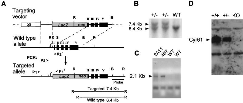
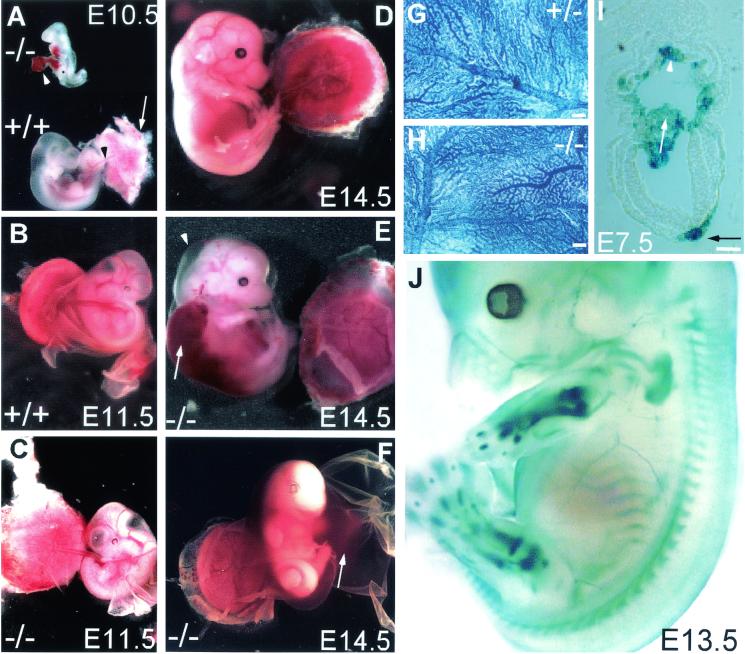
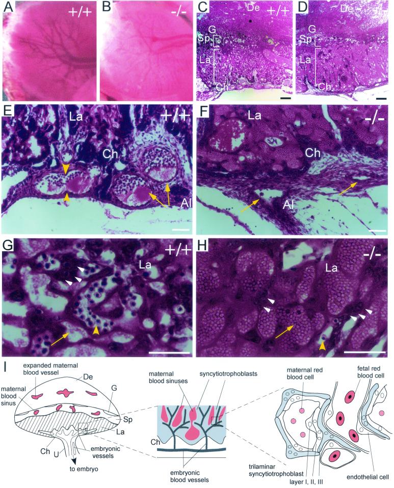
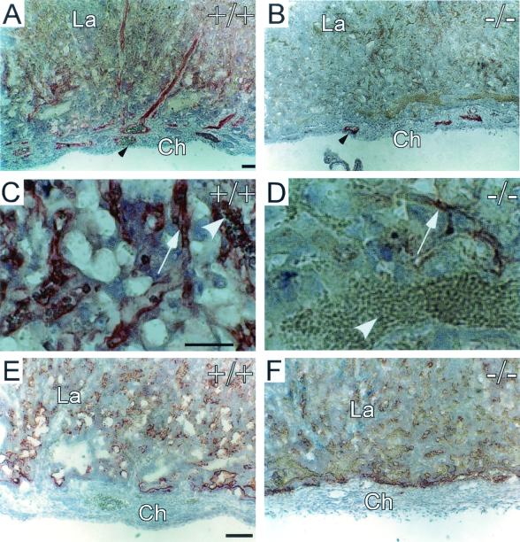
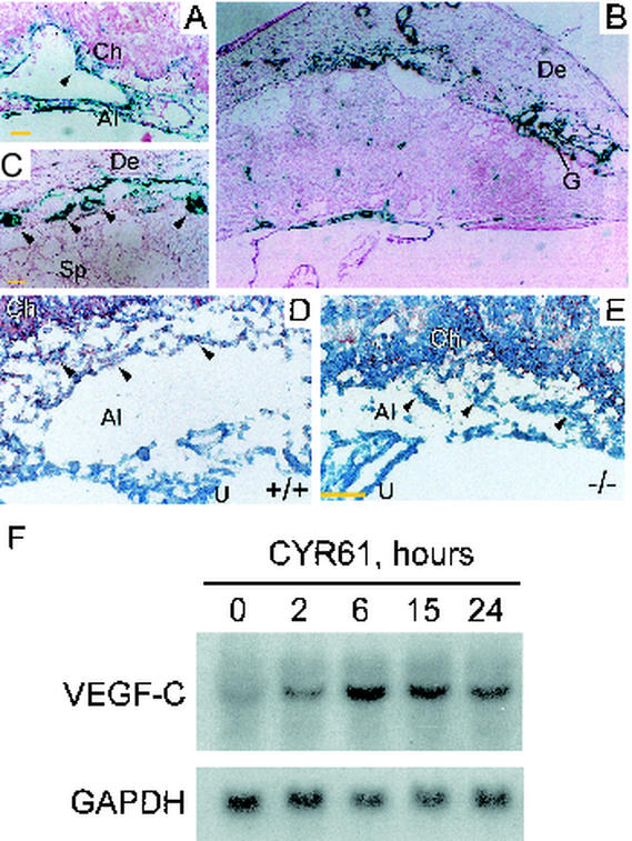
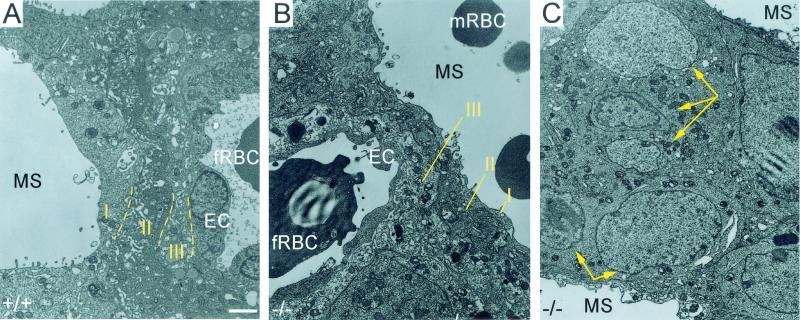
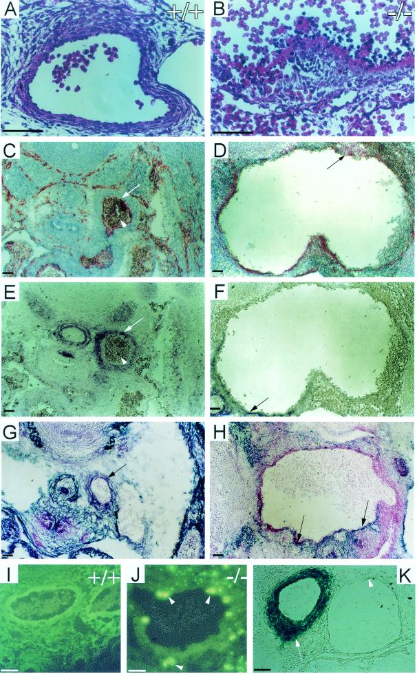
CYR61 (CCN1) is a member of the CCN family of secreted matricellular proteins that includes connective tissue growth factor (CCN2), NOV (CCN3), WISP-1 (CCN4), WISP-2 (CCN5), and WISP-3 (CCN6). First identified as the product of a growth factor-inducible immediate-early gene, CYR61 is an extracellular matrix-associated angiogenic inducer that functions as a ligand of integrin receptors to promote cell adhesion, migration, and proliferation. Aberrant expression of Cyr61 is associated with breast cancer, wound healing, and vascular diseases such as atherosclerosis and restenosis. To understand the functions of CYR61 during development, we have disrupted the Cyr61 gene in mice. We show here that Cyr61-null mice suffer embryonic death: approximately 30% succumbed to a failure in chorioallantoic fusion, and the reminder perished due to placental vascular insufficiency and compromised vessel integrity. These findings establish CYR61 as a novel and essential regulator of vascular development. CYR61 deficiency results in a specific defect in vessel bifurcation (nonsprouting angiogenesis) at the chorioallantoic junction, leading to an undervascularization of the placenta without affecting differentiation of the labyrinthine syncytiotrophoblasts. This unique phenotype is correlated with impaired Vegf-C expression in the allantoic mesoderm, suggesting that CYR61-regulated expression of Vegf-C plays a role in vessel bifurcation. The genetic and molecular basis of vessel bifurcation is presently unknown, and these findings provide new insight into this aspect of angiogenesis.


Figures

FIG. 1.
FIG. 1.
Gene targeting of Cyr61 in mice. (A) Targeting vector. The Cyr61 genomic locus was disrupted by replacement of the XhoI-SmaI fragment, which includes the translation start site, exon I, and half of exon II, with a LacZ-PGK-neo cassette after homologous recombination. B, BamHI; R, EcoRI; S, SmaI; X, XhoI. (B) Southern blot analysis. DNA isolated from ES cell clones was digested with EcoRI and probed with a BamHI-EcoRI fragment (panel A), yielding 6.4- and 7.4-kb bands for the WT and targeted alleles, respectively. (C) PCR. Homologous recombination was confirmed by using a PCR primer pair, P1 and P1′ (panel A), to amplify a 2.1-kb fragment specific to the targeted allele. ES cell clones 2A11 and 4B7 showed the targeted allele. Additionally, PCR primer pairs (P2-P2′ and P2-P1′) yielded a 388-bp fragment and a 600-bp fragment from the WT and targeted alleles, respectively (data not shown). (D) Western blot analysis. Mouse embryo fibroblasts isolated from E11.5 embryos were stimulated with 10% serum for 1 h; total cell lysates were probed with anti-CYR61 antibodies after electrophoresis. KO, knockout.
FIG. 2.
FIG. 2.
Embryonic lethality in Cyr61-deficient mutants. (A) Chorioallantoic fusion occurred in WT embryos by E10.5, with the allantois (black arrowhead) attached to the placenta (white arrow). In some Cyr61−/− embryos, the allantois (white arrowhead) failed to fuse with the placenta, resulting in atrophy. (B) WT embryos at E11.5 showing successful chorioallantoic fusion and healthy placenta. (C) Some Cyr61−/− embryos at E11.5 showing successful chorioallantoic fusion but displaying vascular deficiency in the chorionic plate, resulting in a relatively pale placenta. (D) WT E14.5 embryo. (E) Cyr61−/− embryo (littermate of that shown in panel D) that developed to E14.5, exhibiting edema (arrowhead) and hemorrhage (arrow). (F) Other embryos obtained from E14.5 litters. These embryos were moribund, showing evidence of hemorrhage from the umbilical artery, filling the amnion. Vascular structure appeared normal in hematoxylin-stained yolk sacs from E10.5 Cyr61+/−(G) and Cyr61−/− (H) embryos. Bars in panels G and H, 200 μm. (I) Expression of β-galactosidase driven by the Cyr61 promoter. Expression was detected in both the chorion (white arrowhead) and the allantois (white arrow) at E7.5, when the allantois approached the chorion for eventual fusion. LacZ expression was also detected in the notochordal plate (black arrow). Bar, 10 μm. (J) Whole-mount staining of an E13.5 embryo showing LacZ expression in the sclerotomes of somites, endochondral bones (including those in fore- and hind limbs, digits, and ribs), and large arteries.
FIG. 3.
FIG. 3.
Histological analysis of placental defects in Cyr61−/− embryos. At E12.5, vessel branches had developed in the chorionic plate of the WT (A) but not Cyr61−/− (B) placenta. Hematoxylin and eosin staining revealed that both WT (C) and null (D) placentae developed differentiated zones, including the chorionic plate (Ch), the labyrinth (La), and the spongiotrophoblast layer (Sp) adjacent to the maternal decidua (De). Close-up view (E) shows vessels (arrowheads) undergoing or having completed (arrows) nonsprouting angiogenesis, thus bifurcating the parental vessels at the WT chorionic plate. Al, allantois; G, giant cells. Vessels (arrows) in the Cyr61−/− chorionic plate appear compressed, and no bifurcation was observed (F). Unlike the WT labyrinth (G), where fetal vessels containing nucleated red blood cells (yellow arrowhead) interdigitate with maternal blood sinuses (yellow arrow), the Cyr61−/− labyrinth (H) was predominantly saturated with maternal blood sinuses (arrow) and few fetal vessels were found. Trophoblast nuclei (white arrowheads) were evident in both WT and mutant labyrinth (G and H). A schematic representation of the mouse placenta, with expanded views of the labyrinthine zone, is also shown (I). U, umbilical cord. White bars, 50 μm; black bars in panels C and D, 200 μm.
FIG. 4.
FIG. 4.
Immunostaining of Cyr61−/− placenta. (A) PECAM-1 staining for endothelial cells showed the embryonic vessels (arrowhead) in the WT chorionic plate and labyrinth. (B) Drastically fewer PECAM-staining endothelial cells (arrowhead) were found in the Cyr61−/− placenta. (C) Higher magnification views show that the PECAM-staining fetal vessels (arrow) contained embryonic red blood cells (arrowhead) in the WT labyrinth. (D) In contrast, endothelial cells (arrow) of the Cyr61−/− labyrinth appeared compressed and carried no apparent red blood cells. The white arrowhead in panel D points to maternal blood cells. Despite deficient vascularization of the Cyr61−/− labyrinth, no difference in VEGF-A expression between the WT (E) and Cyr61−/− (F) placentae could be detected by immunostaining. Similar expression of Tie-2 was also observed in WT and Cyr61−/− placentae (data not shown). Ch, chorion, La, labyrinth. Bars, 50 μm.
FIG. 5.
FIG. 5.
(A) β-Galactosidase staining was detected at the chorioallantoic junction at E10.5, when embryonic vessels were actively bifurcating and invaded the placenta. Intense staining was found in the allantoic mesoderm and inner endothelial lining associated with bifurcating vessels (arrowhead). (B) By E13.5, β-galactosidase staining was localized to the vessels in the chorionic plate, maternal decidua, and trophoblast giant cells. (C) Higher magnification of panel B is given to demonstrate staining in giant cells (arrowheads). (D) VEGF-C was detected in the allantoic mesoderm (arrowheads) and chorion of WT E9.5 placenta by immunostaining but not in allantoic mesoderm of Cyr61−/− placenta (E). (F) In Northern blot analysis, serum-starved mouse embryonic fibroblasts isolated from E14.5 WT embryos were treated with purified recombinant CYR61 for various durations before the total RNA was isolated. Vegf-C mRNA was upregulated sixfold after 6 h of CYR61 treatment, and this upregulation was sustained for at least 24 h. The hybridization signal was quantified by Phosphorimager. Al, allantois; Ch, chorion; De, decidua; G, giant cells; La, labyrinth; Sp, spongiotrophoblast; U, umbilical cord. Yellow bars, 50 μm.
FIG. 6.
FIG. 6.
Transmission electron micrographs of the E12.5 labyrinth. Both WT (A) and Cyr61−/− (B) labyrinths displayed the trilaminar barrier composed of two layers of syncytiotrophoblasts (II and III) in addition to a layer of mononuclear trophoblasts (I). In the Cyr61−/− labyrinth, where maternal blood sinuses dominate (C), ectoplacental trophoblasts differentiated and formed syncytium (yellow arrows point to multiple nuclei within fused cells) without neighboring embryonic vessels. EC, endothelial cells; fRBC, fetal red blood cells; mRBC, maternal red blood cells; MS, maternal blood sinuses. White bar, 2 μm.
FIG. 7.
FIG. 7.
Hemorrhage of the dorsal aorta in Cyr61-deficient E13.5 embryos. (A, C, E, G, I) WT embryos. (B, D, F, H, J) Cyr61−/− embryos. Hematoxylin and eosin staining showed completely disorganized vascular cells (B) in the Cyr61−/− dorsal aorta compared to that for the WT (A). Transverse sections were immunostained for PECAM-1 (C and D), α-smooth-muscle actin (E and F), and desmin (G and H), showing the drastically dilated lumen in Cyr61−/− embryos. The endothelial lining (arrows in panels C and D), the smooth-muscle cell wall (arrows in panels E and F), and the pericytes (arrows in panels G and H) of the aorta were disorganized compared to that for the WT, with the apparent mixing of endothelial cells, VSMCs, and pericytes. WT (I) and Cyr61−/− (J) dorsal aorta sections were subjected to TUNEL analysis; apoptotic cells are identified by green fluorescence (arrowheads). β-Galactosidase staining was prominent in arteries such as the dorsal aorta (arrow) but nearly undetectable in the vein (arrowhead) (K). Bars, 50 μm.

Similar articles

Cited by

References

    1. Anson-Cartwright, L., K. Dawson, D. Holmyard, S. J. Fisher, R. A. Lazzarini, and J. C. Cross. 2000. The glial cells missing-1 protein is essential for branching morphogenesis in the chorioallantoic placenta. Nat. Genet. 25:311-314. - PubMed
    1. Babic, A. M., C.-C. Chen, and L. F. Lau. 1999. Fisp12/mouse connective tissue growth factor mediates endothelial cell adhesion and migration through integrin αVβ3, promotes endothelial cell survival, and induces angiogenesis in vivo. Mol. Cell. Biol. 19:2958-2966. - PMC - PubMed
    1. Babic, A. M., M. L. Kireeva, T. V. Kolesnikova, and L. F. Lau. 1998. CYR61, product of a growth factor-inducible immediate-early gene, promotes angiogenesis and tumor growth. Proc. Natl. Acad. Sci. USA 95:6355-6360. - PMC - PubMed
    1. Bader, B. L., H. Rayburn, D. Crowley, and R. O. Hynes. 1998. Extensive vasculogenesis, angiogenesis, and organogenesis precede lethality in mice lacking all alpha v integrins. Cell 95:507-519. - PubMed
    1. Bork, P. 1993. The modular architecture of a new family of growth regulators related to connective tissue growth factor. FEBS Lett. 327:125-130. - PubMed

Publication types

MeSH terms

Substances