

Control of PERK eIF2alpha kinase activity by the endoplasmic reticulum stress-induced molecular chaperone P58IPK
- PMID: 12446838
- PMCID: PMC138540
- DOI: 10.1073/pnas.252341799
Control of PERK eIF2alpha kinase activity by the endoplasmic reticulum stress-induced molecular chaperone P58IPK
Abstract
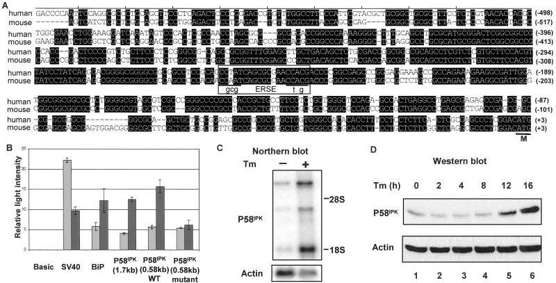
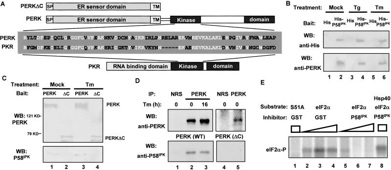
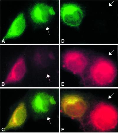
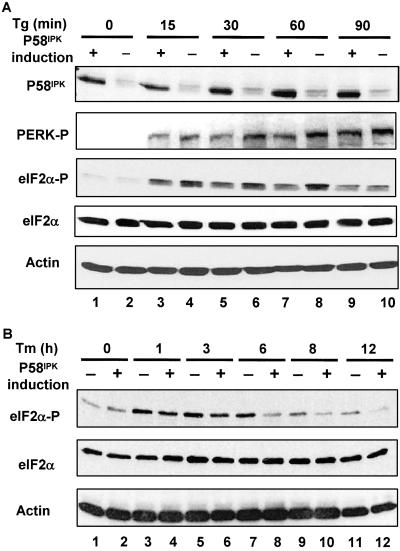
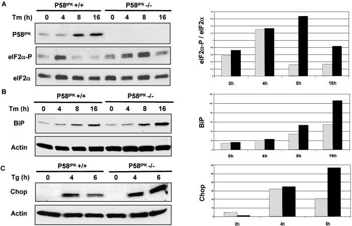
P58(IPK) is an Hsp40 family member known to inhibit the interferon (IFN)-induced, double-stranded RNA-activated, eukaryotic initiation factor 2alpha (eIF2alpha) protein kinase R (PKR) by binding to its kinase domain. We find that the stress of unfolded proteins in the endoplasmic reticulum (ER) activates P58(IPK) gene transcription through an ER stress-response element in its promoter region. P58(IPK) interacts with and inhibits the PKR-like ER-localized eIF2alpha kinase PERK, which is normally activated during the ER-stress response to protect cells from ER stress by attenuating protein synthesis and reducing ER client protein load. Levels of phosphorylated eIF2alpha were lower in ER-stressed P58(IPK)-overexpressing cells and were enhanced in P58(IPK) mutant cells. In the ER-stress response, PKR-like ER kinase (PERK)-mediated translational repression is transient and is followed by translational recovery and enhanced expression of genes that increase the capacity of the ER to process client proteins. The absence of P58(IPK) resulted in increased expression levels of two ER stress-inducible genes, BiP and Chop, consistent with the enhanced eIF2alpha phosphorylation in the P58(IPK) deletion cells. Our studies suggest that P58(IPK) induction during the ER-stress response represses PERK activity and plays a functional role in the expression of downstream markers of PERK activity in the later phase of the ER-stress response.
Figures
Similar articles
-
P58IPK, a novel endoplasmic reticulum stress-inducible protein and potential negative regulator of eIF2alpha signaling.J Biol Chem. 2003 May 2;278(18):15558-64. doi: 10.1074/jbc.M212074200. Epub 2003 Feb 24. J Biol Chem. 2003. PMID: 12601012
-
The cellular protein P58IPK regulates influenza virus mRNA translation and replication through a PKR-mediated mechanism.J Virol. 2007 Mar;81(5):2221-30. doi: 10.1128/JVI.02151-06. Epub 2006 Dec 13. J Virol. 2007. PMID: 17166899 Free PMC article.
-
P58(IPK) inhibition of endoplasmic reticulum stress in human retinal capillary endothelial cells in vitro.Mol Vis. 2008 Jul 13;14:1122-8. Mol Vis. 2008. PMID: 18568130 Free PMC article.
-
[Unfolded protein response: its role in physiology and physiopathology].Med Sci (Paris). 2007 Mar;23(3):291-6. doi: 10.1051/medsci/2007233291. Med Sci (Paris). 2007. PMID: 17349291 Review. French.
-
[Molecular biology of the ER stress response].Seikagaku. 2004 Jul;76(7):617-30. Seikagaku. 2004. PMID: 15346898 Review. Japanese. No abstract available.
Cited by
-
ER residential chaperone GRP78 unconventionally relocalizes to the cell surface via endosomal transport.Cell Mol Life Sci. 2021 Jun;78(12):5179-5195. doi: 10.1007/s00018-021-03849-z. Epub 2021 May 11. Cell Mol Life Sci. 2021. PMID: 33974094 Free PMC article.
-
The microRNA-200 family regulates pancreatic beta cell survival in type 2 diabetes.Nat Med. 2015 Jun;21(6):619-27. doi: 10.1038/nm.3862. Epub 2015 May 18. Nat Med. 2015. PMID: 25985365
-
Lipid Dyshomeostasis and Inherited Cerebellar Ataxia.Mol Neurobiol. 2022 Jun;59(6):3800-3828. doi: 10.1007/s12035-022-02826-2. Epub 2022 Apr 14. Mol Neurobiol. 2022. PMID: 35420383 Free PMC article. Review.
-
Activation of calcium/calmodulin-dependent protein kinase II in obesity mediates suppression of hepatic insulin signaling.Cell Metab. 2013 Dec 3;18(6):803-15. doi: 10.1016/j.cmet.2013.10.011. Epub 2013 Nov 21. Cell Metab. 2013. PMID: 24268736 Free PMC article.
-
Flaccidoxide-13-Acetate-Induced Apoptosis in Human Bladder Cancer Cells is through Activation of p38/JNK, Mitochondrial Dysfunction, and Endoplasmic Reticulum Stress Regulated Pathway.Mar Drugs. 2019 May 13;17(5):287. doi: 10.3390/md17050287. Mar Drugs. 2019. PMID: 31086026 Free PMC article.
References
-
- Kaufman R. J. (2000) in Translational Control of Gene Expression, eds. Sonenberg, N., Hershey, J. W. B. & Mathews, M. B. (Cold Spring Harbor Lab. Press, Plainview, NY), pp. 503–528.
-
- Ron D. & Harding, H. P. (2000) in Translational Control of Gene Expression, eds. Sonenberg, N., Hershey, J. W. B. & Mathews, M. B. (Cold Spring Harbor Lab. Press, Plainview, NY), pp. 547–560.
-
- Lee T. G., Tomita, J., Hovanessian, A. G. & Katze, M. G. (1992) J. Biol. Chem. 267, 14238-14243. - PubMed
Publication types
MeSH terms
Substances
Associated data
- Actions
Grants and funding
LinkOut - more resources
Full Text Sources
Other Literature Sources
Molecular Biology Databases
Research Materials
