. 2002 Dec 24;99(26):17060-5.
doi: 10.1073/pnas.242624699. Epub 2002 Nov 21.

A highly Ca2+-sensitive pool of vesicles is regulated by protein kinase C in adrenal chromaffin cells

Affiliations

A highly Ca2+-sensitive pool of vesicles is regulated by protein kinase C in adrenal chromaffin cells

Yan Yang et al. Proc Natl Acad Sci U S A. .

Abstract

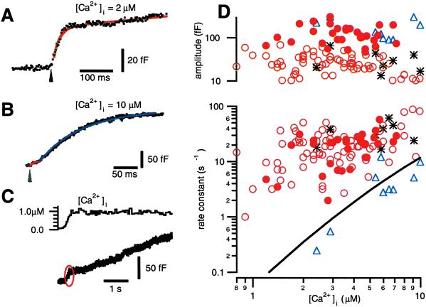
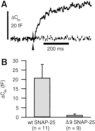
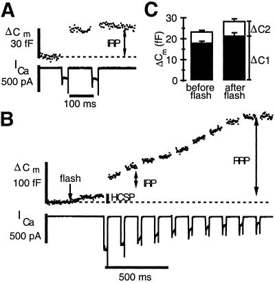
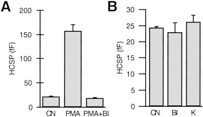
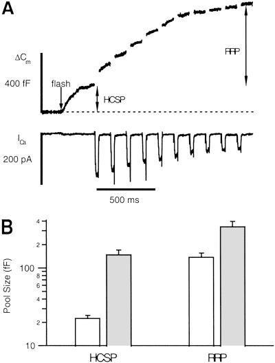
We have used flash photolysis of caged Ca2+ and membrane capacitance measurements to probe exocytosis in chromaffin cells at low concentrations of intracellular Ca2+ ([Ca2+]i) (<10 microM). We observed a small pool of granules that is more sensitive to [Ca2+]i than the previously described "readily releasable pool." Upon activation of PKC, this "highly Ca2+-sensitive pool" is enhanced in size to a greater extent than the readily releasable pool but is eliminated upon expression of a C-terminal deletion mutant (Delta9) of synaptosome-associated protein of 25 kDa (SNAP-25). Thus, in chromaffin cells, PKC enhances exocytosis both by increasing the number of readily releasable vesicles and by shifting vesicles to a highly Ca2+-sensitive state, enabling exocytosis at sites relatively distant from Ca2+ channels.


Figures

Fig 1.
Fig 1.
A small but fast phase of exocytosis is triggered by photoelevation of [Ca2+]i to 1–10 μM. (A) Flash photolysis of caged Ca2+ (arrow) elevates [Ca2+]i to 2.0 μM and elicits a burst of exocytosis with an amplitude of 44 fF and a rate constant of 50 s−1. (B) Flash photolysis elevates [Ca2+]i to 10 μM and elicits exocytosis with two exponential components. The fast component (red) has an amplitude of 17 fF and a rate constant of 140 s−1. The slower component (blue) has an amplitude of 197 fF and a rate constant of 10 s−1. (C) The small but rapid phase of exocytosis (red oval) is also evident when [Ca2+]i is elevated relatively slowly by photolyzing the cage with illumination from a monochromator. (D) The rate constants and amplitudes of exponential fits to the data are plotted vs. the [Ca2+]i that follows flash photolysis of caged Ca2+. Unfilled circles are from single-exponential fits, whereas the triangles and asterisks represent the slow and fast components, respectively, of double exponential fits to the data. The solid line is the rate constant of release for the RRP from the model in ref. . The filled circles represent fits to responses elicited after exposing cells to 100 nM PMA.
Fig 2.
Fig 2.
The C terminus of SNAP-25 is essential for highly Ca2+-sensitive exocytosis. Paired experiments were performed with cells expressing either WT SNAP-25 or SNAP-25 truncated to eliminate the nine C-terminal amino acid residues (Δ9SNAP-25). The Semliki Forest virus expression system was used to express GFP-tagged SNAP-25, and only highly fluorescent cells were selected for experimentation. (A) Sample responses to flash photoelevation of [Ca2+]i from 0.34 to 4.7 μM (dashed line: Δ9SNAP-25) and from 0.40 to 2.7 μM (solid line: WT SNAP-25). (B) Amplitude of highly Ca2+-sensitive responses, defined as the fastest kinetic component of exocytosis. For WT cells, the average [Ca2+]i before and after the flash was 0.53 and 6.0 μM. For Δ9SNAP-25, average [Ca2+]i was elevated from 0.57 to 5.0 μM. Cm, membrane capacitance.
Fig 3.
Fig 3.
The IRP released by membrane depolarization is not highly Ca2+-sensitive. (A) Cm response to a pair of depolarizing pulses from −70 mV to +10 mV, 30 ms in duration. The IRP was allowed to recover for ≈60 s before applying the flash plus depolarization stimuli. (B) Flash photolysis of caged Ca2+ at the time indicated by the arrow elevates [Ca2+]i to 1.8 μM and releases the HCSP. This step was followed 300 ms later by a train of depolarizing pulses to +20 mV. (C) Comparison of the ΔCm resulting from a pair of depolarizing pulses before and after depletion of the HCSP according to the protocol from parts A and B. The filled bars depict the responses to the first pulse, whereas the open bars stacked on top depict the much smaller increase in Cm from the second pulse (n = 16).
Fig 4.
Fig 4.
The size of the HCSP is increased with activation of PKC, but PKC activity is not essential for highly Ca2+-sensitive release. (A) The size of the HCSP is larger after exposure to 100 nM PMA than the control response (CN), but this enhancement is blocked by the PKC inhibitor bisindolylmaleimide I (BI). BI (1 μM) was added to both the bath and pipette solutions 10–60 min before the experiment. n = 8 cells for each condition. (B) Exposure of cells to BI (n = 18) or the nonspecific protein kinase inhibitor K252a (K, 0.5 μM, n = 12) in the absence of PMA has no effect on the size of the HCSP. n = 17 paired CN.
Fig 5.
Fig 5.
The size of the HCSP is increased to a greater extent than the RRP upon activation of PKC. (A) Sample trace after the protocol of Fig. 3B. Flash photoelevation of [Ca2+]i to 4 μM (arrow) reveals a large HCSP. (B) Summary of control responses (open bars, n = 12) and responses in the presence of PMA (filled bars, n = 11). The fractional increase in the HCSP is much greater than the fractional increase in the RRP (note the log scale). The mean [Ca2+]i before photolysis was 0.68 μM for control cells and 0.74 μM for cells in PMA. After photolysis, the mean [Ca2+]i was 3.3 μM for control and 4.1 μM in PMA. Control cells had an initial mean ICa of 294 pA, whereas cells in PMA had a mean initial ICa of 207 pA.

Comment in

Similar articles

Cited by

References

    1. Horrigan F. & Bookman, R. (1994) Neuron 13, 1119-1129. - PubMed
    1. Hsu S. F. & Jackson, M. B. (1996) J. Physiol. (Paris) 494, Part 2, 539-553. - PMC - PubMed
    1. Mennerick S. & Matthews, G. (1996) Neuron 17, 1241-1249. - PubMed
    1. Giovannucci D. R. & Stuenkel, E. L. (1997) J. Physiol. (Paris) 498, Part 3, 735-751. - PMC - PubMed
    1. Moser T. & Neher, E. (1997) J. Neurosci. 17, 2314-2323. - PMC - PubMed

Publication types

LinkOut - more resources