. 2002 Nov;201(5):363-70.
doi: 10.1046/j.0021-8782.2002.00106.x.

Intralobular ducts of human major salivary glands contain leptin and its receptor

Affiliations

Intralobular ducts of human major salivary glands contain leptin and its receptor

R De Matteis et al. J Anat. 2002 Nov.

Abstract

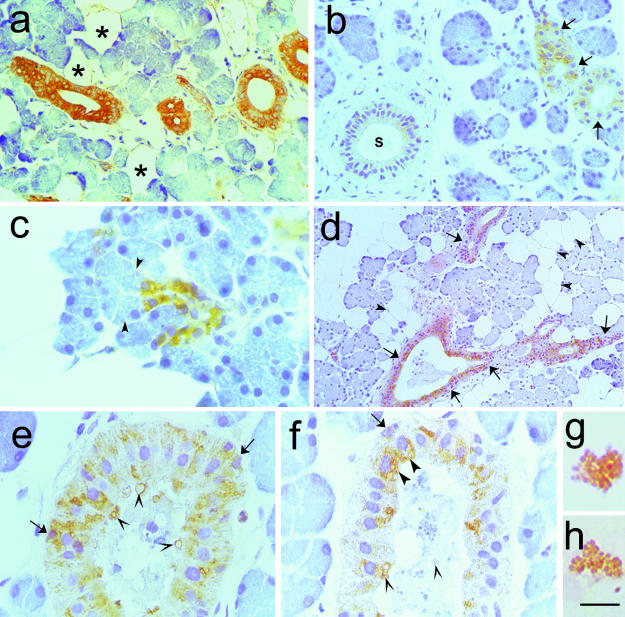
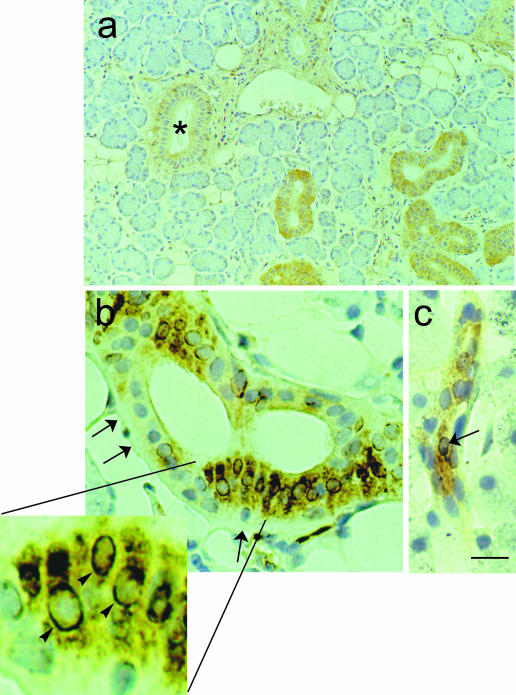
Leptin, a 16-kDa hormone, plays an important role in the control of food intake and in energy homeostasis both in rodents and in man. Leptin is mainly produced and secreted by adipocytes, but other tissues and gastric glands have also recently been shown to produce it in a dual (endocrine and exocrine) mode. In addition, a leptin receptor has been detected in taste cells of mouse circumvallate papillae and in rat intestinal epithelium. These data prompted us to carry out a detailed study of human salivary glands as potential leptin-producing organs. Biopsies of salivary glands (submandibular and parotid) obtained from male and female patients during surgery for different clinical indications were subjected to immunohistochemical study for the presence of leptin, its functional receptor, insulin and glucagon. The presence and cellular distribution of glucocorticoid receptor in leptin-secreting cells were also investigated. Double immunohistochemical staining (silver-gold intensification and avidin-biotin-peroxidase) was used for the visualization of glucocorticoid receptor and leptin labelling, respectively. The results show that intralobular duct cells of submandibular and parotid glands are immunoreactive for leptin, leptin receptor and glucagon but not for insulin. Leptin was also detected in some microglobules in whole saliva obtained from four healthy volunteers. Co-localization for leptin, leptin receptor and glucocorticoid receptor in the same cell type suggested a functional relationship between glucocorticoid hormone and leptin secretion also at the level of the salivary glands.


Figures

Fig. 1
Fig. 1
Immunohistochemistry of human major salivary glands. The primary antibodies were a polyclonal antileptin antiserum (A-20), diluted 1 : 100 (a—c and e—h) and a prediluted polyclonal antiglucagon antiserum (d). Paraffin-embedded tissue, ABC method. (a,b) Intralobular duct epithelial cells showing cytoplasmic staining in both the parotid (a) and the submandibular (b) gland; some completely negative cells were closely apposed to leptin-positive ones (arrows in b). A leptin-negative excretory duct is also visible (S in b). (c) High magnification of leptin-positive parotid tissue. Strongly leptin-stained cuboid cells of a sprouting intercalated duct. The tubuloacinar cells (arrowheads) were not labelled. (d) Glucagon immunoreaction was detected exclusively in the intralobular ducts of a parotid gland. Most epithelial cells were labelled, but negative ones were also visible (arrows). Adipocytes were not labelled (arrowheads). (e,f) At high magnification, leptin immunoreactive granules were evident in the cytoplasm of the epithelial cells in a striated duct (e) and in a distal intercalated (f) duct. Sometimes these leptin-positive granules were in the apical portion of the cell (large arrowheads) or mixed to salivary material (small arrowheads). Some basal cells also stained for leptin (arrows). (g,h) Leptin-labelled microglobules in saliva were observed at high magnification. Scale bars: a,b 7 = 44 μm; c = 17 μm; d = 88 mgr;m; e,f = 20 μm; g,h = 9 μm
Fig. 2
Fig. 2
Immunohistochemistry of human major salivary gland (submandibular). The primary antibody is a polyclonal anti-lepR antiserum (C-20), diluted 1 : 300. Paraffin-embedded tissue, ABC method. The intralobular ducts showed diffuse cytoplasmic staining, whereas the interlobular ducts were not labelled (*). Adipocytes were also lepR-positive. (b,c) Double staining of human major salivary gland (parotid). A silver—gold intensification procedure, modified from the technique described by Danscher et al. (1983), was used for the histochemical visualization of glucocorticoid receptor (GR) antibody—antigen binding (first immunostaining; black). In the second immunostaining, leptin was labelled (yellowish brown) as described above (ABC method). A monoclonal anti-GR antiserum, diluted 1: 20 and a polyclonal antileptin antiserum, diluted 1 : 120, were used. Paraffinembedded tissue. (b) Numerous intralobularduct cells exhibited double (leptin and GR) staining: the cytoplasm, nuclei, or both stained for GR, but the cytoplasm wasalso leptin-positive. Completely negative cells were also present (arrows). High-power inset of the area in (b): some cells showed clear GR staining around the nuclear outline (arrowheads); their cytoplasm was intensely leptin-immunoreactive. (c) Proximal intercalated duct in a different field of the same sample exhibiting leptin-positive cells, one of which also stains for GR (arrow). Scale bars: a = 60 μm; b,c = 12 μm; inset of b = 4.6 μm.

References

    1. Adcock IM, Gilbey T, Gelder CM, Chung KM, Barnes PJ. Glucocorticoids receptor localization in normal and asthmatic lung. Am. J. Respir. Crit. Care Med. 1996;154(3 Part 1):771–782. - PubMed
    1. Antakly T, Eisen HJ. Immunocytochemical localization of glucocorticoid receptor in target cells. Endocrinology. 1984;115:1984–1989. - PubMed
    1. Antakly T, Zhang C, Sarrieau A, Raquidan D. Cell-specific expression of the glucocorticoid receptor within granular convoluted tubules of the rat submaxillary gland. Endocrinology. 1991;128:617–622. - PubMed
    1. Bado A, Levasseur S, Attoub S, Kermorgant S, Laigneau JP, Bortoluzzi MN, et al. The stomach is a source af leptin. Nature. 1998;394:790–793. - PubMed
    1. Barka T. Biologically active polypeptides in submandibular glands. J. Histochem. Cytochem. 1980;28:836–859. - PubMed

Publication types

LinkOut - more resources