

Analysis of baseline and cisplatin-inducible gene expression in Fanconi anemia cells using oligonucleotide-based microarrays
- PMID: 12450415
- PMCID: PMC138804
- DOI: 10.1186/1471-2326-2-5
Analysis of baseline and cisplatin-inducible gene expression in Fanconi anemia cells using oligonucleotide-based microarrays
Abstract
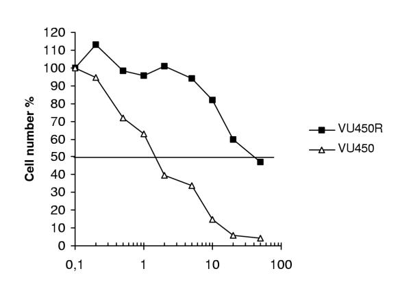
BACKGROUND: Patients with Fanconi anemia (FA) suffer from multiple defects, most notably of the hematological compartment (bone marrow failure), and susceptibility to cancer. Cells from FA patients show increased spontaneous chromosomal damage, which is aggravated by exposure to low concentrations of DNA cross-linking agents such as mitomycin C or cisplatin. Five of the identified FA proteins form a nuclear core complex. However, the molecular function of these proteins remains obscure. METHODS: Oligonucleotide microarrays were used to compare the expression of approximately 12,000 genes from FA cells with matched controls. Expression profiles were studied in lymphoblastoid cell lines derived from three different FA patients, one from the FA-A and two from the FA-C complementation groups. The isogenic control cell lines were obtained by either transfecting the cells with vectors expressing the complementing cDNAs or by using a spontaneous revertant cell line derived from the same patient. In addition, we analyzed expression profiles from two cell line couples at several time points after a 1-hour pulse treatment with a discriminating dose of cisplatin. RESULTS: Analysis of the expression profiles showed differences in expression of a number of genes, many of which have unknown function or are difficult to relate to the FA defect. However, from a selected number of proteins involved in cell cycle regulation, DNA repair and chromatin structure, Western blot analysis showed that p21waf1/Cip1 was significantly upregulated after low dose cisplatin treatment in FA cells specifically (as well as being expressed at elevated levels in untreated FA cells). CONCLUSIONS: The observed increase in expression of p21waf1/Cip1 after treatment of FA cells with crosslinkers suggests that the sustained elevated levels of p21waf1/Cip1 in untreated FA cells detected by Western blot analysis likely reflect increased spontaneous damage in these cells.
Figures


Similar articles
-
Differential expression of TP53 associated genes in Fanconi anemia cells after mitomycin C and hydroxyurea treatment.Mutat Res. 2008 Oct 30;656(1-2):1-7. doi: 10.1016/j.mrgentox.2008.06.012. Epub 2008 Jul 5. Mutat Res. 2008. PMID: 18647660
-
Function of the Fanconi anemia pathway in Fanconi anemia complementation group F and D1 cells.Exp Hematol. 2001 Dec;29(12):1448-55. doi: 10.1016/s0301-472x(01)00754-8. Exp Hematol. 2001. PMID: 11750104
-
Phenotypic correction of Fanconi anemia in human hematopoietic cells with a recombinant adeno-associated virus vector.J Clin Invest. 1994 Oct;94(4):1440-8. doi: 10.1172/JCI117481. J Clin Invest. 1994. PMID: 7929819 Free PMC article.
-
Current knowledge on the pathophysiology of Fanconi anemia: from genes to phenotypes.Int J Hematol. 2001 Jul;74(1):33-41. doi: 10.1007/BF02982547. Int J Hematol. 2001. PMID: 11530803 Review.
-
Fanconi anemia and ubiquitination.J Genet Genomics. 2007 Jul;34(7):573-80. doi: 10.1016/S1673-8527(07)60065-4. J Genet Genomics. 2007. PMID: 17643942 Review.
Cited by
-
Differential gene expression in normal human mammary epithelial cells treated with malathion monitored by DNA microarrays.Environ Health Perspect. 2005 Aug;113(8):1046-51. doi: 10.1289/ehp.7311. Environ Health Perspect. 2005. PMID: 16079077 Free PMC article.
-
Oxidative stress-related mechanisms are associated with xenobiotics exerting excess toxicity to Fanconi anemia cells.Environ Health Perspect. 2003 Nov;111(14):1699-703. doi: 10.1289/ehp.6229. Environ Health Perspect. 2003. PMID: 14594617 Free PMC article.
References
-
- Auerbach AD, Buchwald M, Joenje H. Fanconi Anemia. In: Vogelstein B, Kinzler KW, editor. The Genetic Basis of Human Cancer. New York, McGraw-Hill; 1998. pp. 317–332.
-
- The Fanconi anaemia/breast cancer consortium Positional cloning of the Fanconi anaemia group A gene. Nat Genet. 1996;14:324–328. - PubMed
-
- Lo ten Foe JR, Rooimans MA, Bosnoyan-Collins L, Alon N, Wijker M, Parker L, Lightfoot J, Carreau M, Callen DF, Savoia A, et al. Expression cloning of a cDNA for the major Fanconi anaemia gene, FAA. Nat Genet. 1996;14:320–323. - PubMed
LinkOut - more resources
Full Text Sources
Miscellaneous
