. 2002 Dec;68(12):5877-81.
doi: 10.1128/AEM.68.12.5877-5881.2002.

Identification of an iron-regulated, hemin-binding outer membrane protein in Sinorhizobium meliloti

Affiliations

Identification of an iron-regulated, hemin-binding outer membrane protein in Sinorhizobium meliloti

Federico Battistoni et al. Appl Environ Microbiol. 2002 Dec.

Abstract

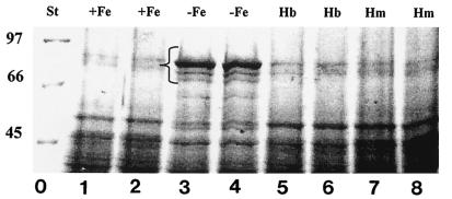
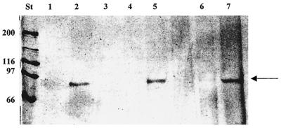
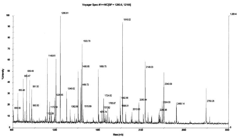
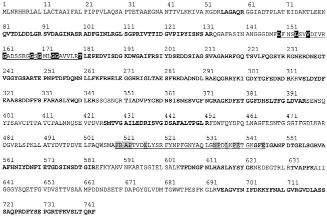
Rhizobia are soil bacteria that are able to establish symbiotic associations with leguminous hosts. In iron-limited environments these bacteria can use iron present in heme or heme compounds (hemoglobin, leghemoglobin). Here we report the presence in Sinorhizobium meliloti of an iron-regulated outer membrane protein that is able to bind hemin but not hemoglobin. Protein assignment was done by matrix-assisted laser desorption ionization-time of flight mass spectrometry. Tryptic peptides correlated with the mass measurements obtained accounted for 54% of the translated sequence of a putative heme receptor gene present in the chromosome of S. meliloti 1021. The results which we obtained suggest that this protein (designated ShmR for Sinorhizobium heme receptor) is involved in high-affinity heme-mediated iron transport.


Figures

FIG. 1.
FIG. 1.
SDS-PAGE analysis of S. meliloti 242 outer membrane proteins. S. meliloti 242 was grown in M3 medium containing 37 μM FeCl3 (lanes 1 and 2), 500 μM EDDHA (lanes 3 and 4), 500 μM EDDHA and 4 μM hemoglobin (lanes 5 and 6), or 500 μM EDDHA and 16 μM hemin (lanes 7 and 8). Outer membrane proteins were extracted as described in the text, electrophoresed in a 10% acrylamide gel, and stained with 0.1% Coomassie brilliant blue. The bracket indicates the position of IROMPs. The positions of molecular mass standards (in kilodaltons) (lane 0) are indicated on the left. St, standards; Hb, hemoglobin; Hm, hemin.
FIG. 2.
FIG. 2.
Binding of the 91-kDa protein to hemin-agarose. Outer membrane proteins were isolated from bacteria grown in M3 medium containing 37 μM FeCl3 (lane 1), 500 μM EDDHA (lane 2), 500 μM EDDHA and 4 μM hemoglobin (lane 3), 500 μM EDDHA and 16 μM hemin (lane 4), or 16 μM PPIX (lane 5), and they were added to hemin-agarose resin. Lanes 6 and 7 contained the hemin-binding proteins from competition assays performed with hemin (lane 6) and PPIX (lane 7). Hemin-binding proteins were eluted as described in Materials and Methods, separated by SDS-PAGE, and silver stained. The arrow indicates the position of the 91-kDa hemin-binding outer membrane protein. The positions of molecular mass standards (in kilodaltons) (lane St) are indicated on the left.
FIG. 3.
FIG. 3.
Mass spectrum obtained by MALDI-TOF mass spectrometry by using the 91-kDa outer membrane protein in a gel digested with trypsin.
FIG. 4.
FIG. 4.
Amino acid sequence of the putative iron transport protein from S. meliloti 1021 (ShmR). Boldface type indicates the experimental peptides identified by MALDI-TOF mass spectrometry. A probable TonB box (residues 149 to 180) and the FRAP-NPNL motif of heme receptors (residues 509 to 545) are underlined. Highly conserved residues present in the TonB box are indicated by a black background, whereas highly conserved residues of the FRAP-NPNL region are indicated by a grey background. The conserved histidine between the FRAP and NPNL motifs is probably replaced by an asparagine (residue 523) in ShmR.

References

    1. Bracken, C. S., M. T. Baer, A. Abdur-Rashid, W. Helmes, and I. Stojiljkovic. 1999. Use of heme-protein complex by the Yersinia enterocolitica HemR receptor: histidine residues are essential for receptor function. J. Bacteriol. 181:6063-6072. - PMC - PubMed
    1. Capela, D., F. Barloy-Hubler, J. Gouzy, G. Bothe, F. Ampe, J. Batut, P. Boistard, A. Becker, M. Boutry, E. Cadieu, S. Dréano, S. Gloux, T. Godrie, A. Goffeau, D. Kahn, E. Kiss, V. Lelaure, D. Masuy, T. Pohl, D. Portetelle, A. Pühler, B. Purnelle, U. Ramsperger, C. Renard, P. Thébault, M. Vandenbol, S. Weidner, and F. Galibert. 2001. Analysis of the chromosome sequence of the legume symbiont Sinorhizobium meliloti strain 1021. Proc. Natl. Acad. Sci. USA 98:9877-9882. - PMC - PubMed
    1. Carson, K. C., A. R. Glenn, and M. J. Dilworth. 1994. Specificity of siderophore-mediated transport of iron in rhizobia. Arch. Microbiol. 161:333-339.
    1. Dashper, S. G., A. Hendtlass, N. Slakeski, C. Jackson, K. J. Croos, L. Brownfield, R. Hamilton, I. Barr, and E. C. Reynolds. 2000. Characterization of a novel outer membrane hemin-binding protein of Porphyromonas gingivalis. J. Bacteriol. 182:6456-6462. - PMC - PubMed
    1. Fabiano, E., G. Gualtieri, C. Pritch, G. Polla, and A. Arias. 1994. Extent of high-affinity iron transport systems in field isolates of rhizobia. Plant Soil 164:177-185.

Publication types

MeSH terms