

Surface display of foreign epitopes on the Lactobacillus brevis S-layer
- PMID: 12450814
- PMCID: PMC134443
- DOI: 10.1128/AEM.68.12.5943-5951.2002
Surface display of foreign epitopes on the Lactobacillus brevis S-layer
Abstract
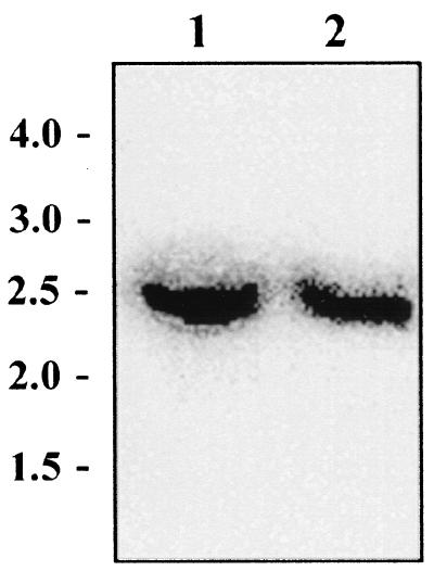
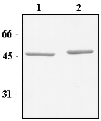
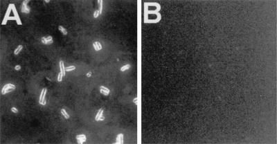
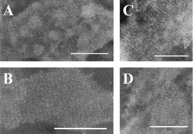
So far, the inability to establish viable Lactobacillus surface layer (S-layer) null mutants has hampered the biotechnological applications of Lactobacillus S-layers. In this study, we demonstrate the utilization of Lactobacillus brevis S-layer subunits (SlpA) for the surface display of foreign antigenic epitopes. With an inducible expression system, L. brevis strains producing chimeric S-layers were obtained after testing of four insertion sites in the slpA gene for poliovirus epitope VP1, that comprises 10 amino acids. The epitope insertion site allowing the best surface expression was used for the construction of an integration vector carrying the gene region encoding the c-Myc epitopes from the human c-myc proto-oncogene, which is composed of 11 amino acids. A gene replacement system was optimized for L. brevis and used for the replacement of the wild-type slpA gene with the slpA-c-myc construct. A uniform S-layer, displaying on its surface the desired antigen in all of the S-layer protein subunits, was obtained. The success of the gene replacement and expression of the uniform SlpA-c-Myc recombinant S-layer was confirmed by PCR, Southern blotting MALDI-TOF mass spectrometry, whole-cell enzyme-linked immunosorbent assay, and immunofluorescence microscopy. Furthermore, the integrity of the recombinant S-layer was studied by electron microscopy, which indicated that the S-layer lattice structure was not affected by the presence of c-Myc epitopes. To our knowledge, this is the first successful expression of foreign epitopes in every S-layer subunit of a Lactobacillus S-layer while still maintaining the S-layer lattice structure.
Figures


References
-
- Bhowmik, T., and J. L. Steele. 1993. Development of an electroporation procedure for gene disruption in Lactobacillus helveticus CNRZ32. J. Gen. Microbiol. 139:1-7.
-
- Bingle, W. H., J. F. Nomellini, and J. Smit. 1997. Cell-surface display of a Pseudomonas aeruginosa strain K pilin peptide within the paracrystalline S-layer of Caulobacter crescentus. Mol. Microbiol. 26:277-288. - PubMed
-
- Boot, H. J., and P. H. Pouwels. 1996. Expression, secretion and antigenic variation of bacterial S-layer proteins. Mol. Microbiol. 21:1117-1123. - PubMed
-
- Breitwieser, A., E. M. Egelseer, D. Moll, N. Ilk, C. Hotzy, B. Bohle, C. Ebner, U. B. Sleytr, and M. Sára. 2002. A recombinant bacterial cell surface (c-layer)-major birch pollen allergen-fusion protein (rSbsC/Bet v1) maintains the ability to self- assemble into regularly structured monomolecular lattices and the functionality of the allergen. Protein Eng. 15:243-249. - PubMed
Publication types
MeSH terms
Substances
LinkOut - more resources
Full Text Sources
Other Literature Sources
