. 2002 Dec;68(12):6043-50.
doi: 10.1128/AEM.68.12.6043-6050.2002.

The genus Caedibacter comprises endosymbionts of Paramecium spp. related to the Rickettsiales (Alphaproteobacteria) and to Francisella tularensis (Gammaproteobacteria)

Affiliations

The genus Caedibacter comprises endosymbionts of Paramecium spp. related to the Rickettsiales (Alphaproteobacteria) and to Francisella tularensis (Gammaproteobacteria)

Cora L Beier et al. Appl Environ Microbiol. 2002 Dec.

Abstract

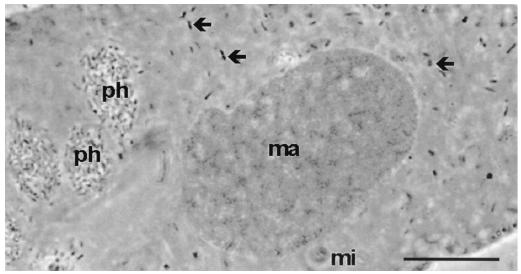
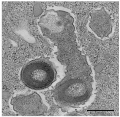
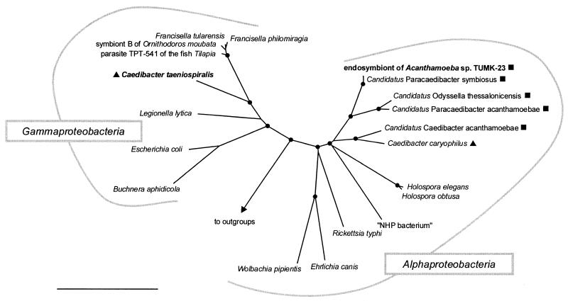
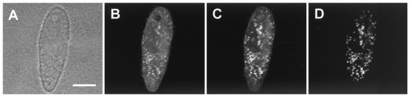
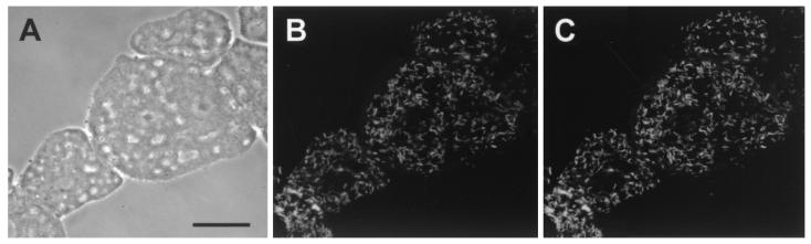
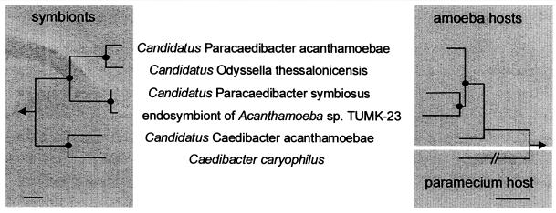
Obligate bacterial endosymbionts of paramecia able to form refractile inclusion bodies (R bodies), thereby conferring a killer trait upon their ciliate hosts, have traditionally been grouped into the genus CAEDIBACTER: Of the six species described to date, only the Paramecium caudatum symbiont Caedibacter caryophilus has been phylogenetically characterized by its 16S rRNA gene sequence, and it was found to be a member of the Alphaproteobacteria related to the RICKETTSIALES: In this study, the Caedibacter taeniospiralis type strain, an R-body-producing cytoplasmatic symbiont of Paramecium tetraurelia strain 51k, was investigated by comparative 16S rRNA sequence analysis and fluorescence in situ hybridization with specific oligonucleotide probes. C. taeniospiralis is not closely related to C. caryophilus (80% 16S rRNA sequence similarity) but forms a novel evolutionary lineage within the Gammaproteobacteria with the family Francisellaceae as a sister group (87% 16S rRNA sequence similarity). These findings demonstrate that the genus Caedibacter is polyphyletic and comprises at least two phylogenetically different bacterial species belonging to two different classes of the PROTEOBACTERIA: Comparative phylogenetic analysis of C. caryophilus, five closely related Acanthamoeba endosymbionts (including one previously uncharacterized amoebal symbiont identified in this study), and their hosts suggests that the progenitor of the alphaproteobacterial C. caryophilus lived within acanthamoebae prior to the infection of paramecia.


Figures

FIG. 1.
FIG. 1.
Phase-contrast micrograph of P. tetraurelia strain 51k stained with orcein. Numerous symbionts (arrows) in the cytoplasm and bacteria in phagosomes (ph) are visible. ma, macronucleus; mi, micronucleus. Bar, 10 μm.
FIG. 2.
FIG. 2.
Ultrastructure of C. taeniospiralis harboring type 51 R bodies within the cytoplasm of its host, P. tetraurelia. Bar, 0.5 μm.
FIG. 3.
FIG. 3.
16S rRNA-based neighbor-joining tree showing (i) the phylogenetic affiliation of C. taeniospiralis 51k (endosymbiont of P. tetraurelia) with representative members of the Gammaproteobacteria and (ii) the relationship of the endosymbiont of Acanthamoeba sp. strain TUMK-23 with the alphaproteobacterial C. caryophilus (endosymbiont of P. caudatum) and other representative members of the Alphaproteobacteria.Candidatus Caedibacter acanthamoebae,” “Candidatus Paracaedibacter acanthamoebae,” “Candidatus Paracaedibacter symbiosus,” and “Candidatus Odyssella thessalonicensis” were recently described as endosymbionts of acanthamoebae (5, 18). The respective eukaryotic hosts are indicated by symbols: ▴, Paramecium sp.; ▪, Acanthamoeba sp. Parsimony bootstrap values (1,000 resamplings) of >97% are indicated as solid circles. “NHP bacterium,” shrimp pathogen causing necrotizing hepatopancreatitis. Bar, 10% estimated evolutionary distance.
FIG. 4.
FIG. 4.
In situ identification of C. taeniospiralis 51k within its host, P. tetraurelia. Identical microscopic fields are depicted. The fluorescence images (B to D) show a median section through the paramecium cell seen in the simulated phase-contrast image (A). Shown is FISH using the FLUOS-labeled oligonucleotide probe EUB338 (B), the Cy3-labeled endosymbiont-specific probe S-S-Ctaenio-129-a-A-18 (C), and the Cy5-labeled endosymbiont-specific probe S-S-Ctaenio-998-a-A-18 (D). Bar, 20 μm
FIG. 5.
FIG. 5.
In situ identification of the bacterial endosymbiont of Acanthamoeba sp. strain TUMK-23. Identical microscopic fields are depicted. (A) Phase-contrast image. (B and C) FISH using FLUOS-labeled oligonucleotide probe EUB338 (B) and Cy3-labeled C. caryophilus-specific probe CC23a (C). Bar, 10 μm.
FIG. 6.
FIG. 6.
Ultrastructure of the C. caryophilus-related endosymbiont (solid arrows) within the cytoplasm of its host, Acanthamoeba sp. strain TUMK-23. cm, amoebal cell membrane; n, nucleus. The open arrows indicate mitochondria. Bar, 0.5 μm.
FIG. 7.
FIG. 7.
Comparison of 16S and 18S rRNA-based neighbor-joining dendrograms of alphaproteobacterial C. caryophilus-related endosymbionts (left) and their Acanthamoeba or Paramecium host organisms (right) suggesting (i) coevolution between bacterial symbionts and Acanthamoeba hosts and (ii) relatively recent transfer of a C. caryophilus-like symbiont from Acanthamoeba to Paramecium. The 18S rRNA gene sequence of the amoebal host of “Candidatus Odyssella thessalonicensis” is not available (6). Parsimony bootstrap values (1,000 resamplings) higher than 99% are indicated as solid circles. The arrow points to outgroups. Bars, 10% estimated evolutionary distance.

Similar articles

Cited by

References

    1. Alm, E. W., D. B. Oerther, M. Larsen, D. A. Stahl, and L. Raskin. 1996. The oligonucleotide probe database. Appl. Environ. Microbiol. 62:3557-3559. - PMC - PubMed
    1. Altschul, S. F., T. L. Madden, A. A. Schaffer, J. Zhang, Z. Zhang, W. Miller, and D. J. Lipman. 1997. Gapped BLAST and PSI-BLAST: a new generation of protein database search programs. Nucleic Acids Res. 25:3389-3402. - PMC - PubMed
    1. Amann, R., N. Springer, W. Ludwig, H. D. Görtz, and K. H. Schleifer. 1991. Identification in situ and phylogeny of uncultured bacterial endosymbionts. Nature 351:161-164. - PubMed
    1. Beale, G. H., and A. Jurand. 1966. Three different types of mate-killer (mu) particles in Paramecium aurelia, stock 540. J. Gen. Microbiol. 23:243-252. - PubMed
    1. Beale, G. H., A. Jurand, and J. R. Preer. 1969. The classes of endosymbionts of Paramecium aurelia. J. Cell Sci. 5:65-91. - PubMed

Publication types

Substances

LinkOut - more resources