. 2002 Dec;68(12):6292-9.
doi: 10.1128/AEM.68.12.6292-6299.2002.

Coexistence of multiple proteobacterial endosymbionts in the gills of the wood-boring Bivalve Lyrodus pedicellatus (Bivalvia: Teredinidae)

Affiliations

Coexistence of multiple proteobacterial endosymbionts in the gills of the wood-boring Bivalve Lyrodus pedicellatus (Bivalvia: Teredinidae)

Daniel L Distel et al. Appl Environ Microbiol. 2002 Dec.

Abstract

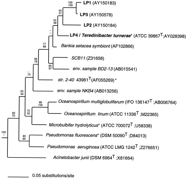
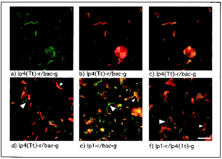
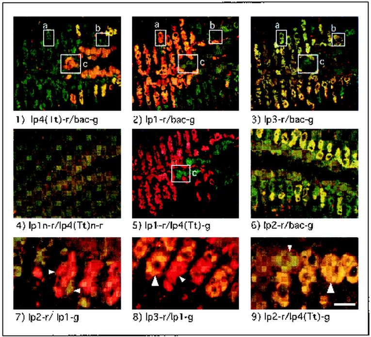
Wood-boring bivalves of the family Teredinidae (commonly called shipworms) are known to harbor dense populations of gram-negative bacteria within specialized cells (bacteriocytes) in their gills. These symbionts are thought to provide enzymes, e.g., cellulase and dinitrogenase, which assist the host in utilizing wood as a primary food source. A cellulolytic, dinitrogen-fixing bacterium, Teredinibacter turnerae, has been isolated from the gill tissues of numerous teredinid bivalves and has been proposed to constitute the sole or predominant symbiont of this bivalve family. Here we demonstrate that one teredinid species, Lyrodus pedicellatus, contains at least four distinct bacterial 16S rRNA types within its gill bacteriocytes, one of which is identical to that of T. turnerae. Phylogenetic analyses indicate that the three newly detected ribotypes are derived from gamma proteobacteria that are related to but distinct (>6.5% sequence divergence) from T. turnerae. In situ hybridizations with 16S rRNA-directed probes demonstrated that the pattern of occurrence of symbiont ribotypes within bacteriocytes was predictable and specific, with some bacteriocytes containing two symbiont ribotypes. However, only two of the six possible pairwise combinations of the four ribotypes were observed to cooccur within the same host cells. The results presented here are consistent with the existence of a complex multiple symbiosis in this shipworm species.


Figures

FIG. 1.
FIG. 1.
A phylogenetic tree for T. turnerae, ribotypes LP1 through LP4, and representative gamma proteobacteria. The tree shown is extracted from a single best tree inferred by maximum likelihood analysis with 16S rRNA sequences. The maximum likelihood tree is identical to one of four best trees inferred by maximum parsimony. Bootstrap proportions are indicated at each node (maximum likelihood above, maximum parsimony below) as a percentage of 1,000 replicates. Dashes indicate bootstrap proportions of <50%. Type cultures and taxa with known cellulolytic capabilities are indicated by the superscripts T and *, respectively. Culture collection and sequence database accession numbers are in given in parentheses. Taxa included in the analyses but not shown in the tree are Aeromonas salmonicida (CCM 4103T; AT009859), Bathymodiolus puteoserpentis chemoautotrophic symbiont (U29163), Bathymodiolus puteoserpentis methanotrophic symbiont (U29164), Erwinia carotovora* (ATCC 15713T; M59149), Lucina nassula symbiont (X95299), Serratia marcescens* (ATCC 13880T; M59160), Vibrio harveyi (ATCC 14126T; X56578), Xanthomonas campestris* (LMG 568T; X95917), and Xanthomonas albilinians* (LMG 494T; X95918).
FIG. 2.
FIG. 2.
Dual-probe in situ hybridizations with ribotype-specific and bacterial-domain-specific probes to label individual bacterial cells from T. turnerae pure culture and L. pedicellatus gill homogenate. Pure-cultured T. turnerae (a-c) and symbionts from L. pedicellatus gill homogenate (d-f) stained with probe combinations indicated below the corresponding panel and viewed with the green (a), red (b), or red and green dual-wavelength filter sets (c-f) are shown. Pure cultures of the symbiont T. turnerae stained uniformly with ribotype- and domain-specific probes, while symbionts in shipworm gill homogenate were heterogeneous with respect to staining with ribotype-specific probes. Large arrows indicate cells stained green by a single domain-specific (d and e) or ribotype-specific (f) probe, while small arrows indicate cells stained yellow to orange due to the combination of red- and green-labeled probes (d and e) or red due to staining with a single ribotype-specific probe (f). Similar results were observed for probe combinations lp3-r-lp4(Tt)-g and lp3-r-bac-g (data not shown). Scale bar, 5 μm.
FIG. 3.
FIG. 3.
Dual-probe in situ hybridizations with ribotype-specific and bacteria-domain-specific probes to label symbiont cells in sectioned gill tissue from L. pedicellatus. Panels 1 to 5 show consecutive sections from a single specimen of L. pedicellatus. Probe combinations applied to each section are indicated below the corresponding panel. Boxes a to c delineate bacteriocytes that could be identified by size, shape, and position in several sections. Small arrows in panels 7 to 9 delineate individual bacteriocytes stained by a single red or green ribotype-specific probe; large arrows delineate yellow- to orange-stained bacteriocytes, indicating the colocalization of two ribotype-specific probes. Note that symbiont ribotypes LP1 and LP3 are localized in a different subset of bacteriocytes than ribotypes LP2 and LP4. Scale bar, 75 μm (panels 1 to 6) or 25 μm (panels 7 to 9).

References

    1. Amann, G., K. O. Stetter, E. Llobet-Brossa, R. Amann, and J. Anton. 2000. Direct proof for the presence and expression of two 5% different 16S rRNA genes in individual cells of Haloarcula marismortui. Extremophiles 4:373-376. - PubMed
    1. Amann, R. I., L. Krumholz, and D. A. Stahl. 1990. Fluorescent-oligonucleotide probing of whole cells for determinative, phylogenetic, and environmental studies in microbiology. J. Bacteriol. 172:762-770. - PMC - PubMed
    1. Clayton, R. A., G. Sutton, P. S. Hinkle, Jr., C. Bult, and C. Fields. 1995. Intraspecific variation in small-subunit rRNA sequences in GenBank: why single sequences may not adequately represent prokaryotic taxa. Int. J. Syst. Bacteriol. 45:595-599. - PubMed
    1. Coughlan, M. P., and F. Mayer. 1992. The cellulose-decomposing bacteria and their enzyme systems, p. 451-516. In A. Ballows, H. Truper, W. Harder, and K. H. Schleifer (ed.), vol. 4. Springer-Verlag, Berlin, Germany.
    1. DeLong, E. F., G. S. Wickham, and N. R. Pace. 1989. Phylogenetic stains: ribosomal RNA-based probes for the identification of single cells. Science 243:1360-1363. - PubMed

Publication types

Substances

LinkOut - more resources