

Defining the caudal ventral striatum in primates: cellular and histochemical features
- PMID: 12451107
- PMCID: PMC2481229
- DOI: 10.1523/JNEUROSCI.22-23-10078.2002
Defining the caudal ventral striatum in primates: cellular and histochemical features
Abstract
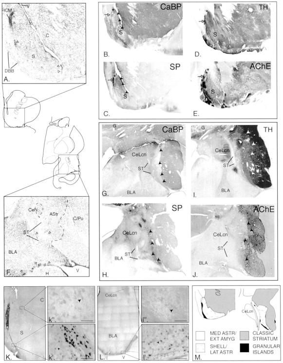
Afferents from the amygdala help to define the ventral striatum and mediate goal-directed behaviors. In addition to well known inputs to the classic ventral striatum, the amygdala also projects to the caudoventral striatum and amygdalostriatal area. We examined whether the primate caudoventral striatum and amygdalostriatal area can be considered part of the "ventral" striatum based on cellular and histochemical features found in the classic rostral ventral striatum. We used several histochemical stains, including calbindin-D28k, a marker of the shell compartment, acetylcholinesterase, substance P, tyrosine hydroxylase, and Bcl-2, a marker of immature neurons, to examine this question. Our results indicate that the lateral amygdalostriatal area and caudoventral striatum are "striatal like" based on intermediate to high acetylcholinesterase and tyrosine hydroxylase levels. The lateral amygdalostriatal area is chemically similar to the shell, whereas the caudoventral striatum more closely resembles the striatum outside the shell. In contrast, the medial amygdalostriatal area is more related to the central amygdaloid nucleus than to the striatum. Bcl-2 immunoreactivity is associated with granular islands and medium-sized cells in the vicinity of the ventral striatum both rostrally and caudally. Together, the caudal ventral striatum has a histochemical and cellular organization similar to that of the rostral ventral striatum, consistent with their common innervation by the amygdala and other ventral structures. In addition, Bcl-2 is expressed in and near both poles of the ventral striatum, suggesting that these areas maintain a heightened capacity for growth and plasticity compared with other striatal sectors.
Figures
References
-
- Bassareo V, Di Chiara G. Differential responsiveness of dopamine transmission to food-stimuli in nucleus accumbens shell/core compartments. Neuroscience. 1999;89:637–642. - PubMed
-
- Breiter HC, Aharon I, Kahneman D, Dale A, Shizgal P. Functional imaging of neural responses to expectancy and experience of monetary gains and losses. Neuron. 2001;30:619–639. - PubMed
Publication types
MeSH terms
Substances
Grants and funding
LinkOut - more resources
Full Text Sources